Wednesday, July 30, 2014

Viral infections and the kidney

Hepatitis C and the kidney

Thanks to Dr. David Frost and Team 1 for presenting an interesting case in morning report today:

We discussed hepatitis C, and the various mechanisms of acute kidney injury specific to the infection. 

Our case had a sub-acute course of 6 weeks duration in which the creatinine increased from a baseline of 69 to 677 on the day of presentation.

We also discussed the importance of the physical exam, making sure to search for findings suggestive of fluid overload, which may push you towards dialysis, and also looking for specific findings of cryoglobulinemia (palpable purpura).  In any patient with acute renal failure, searching for findings that might lead to urgent dialysis is of high priority and so listening for pericardial rubs, and looking for signs of uremic encephalopathy (asterixis, decreased LOC) is important.

Investigations:

The initial investigations showed a normocytic anemia.  There was a non-anion gap metabolic acidosis with a normal potassium.  The liver enzymes and function tests were normal.  The blood film, looking for findings of HHS or TTP, did not show any schistocytes.

The urinalysis, also known as the physical exam of the kidney, showed 3+ protein, trace blood, and the microscopy showed heme-granular casts.  The urine sodium was 24.  The abdominal ultrasound did not show any hydronephrosis or ascites, and the kidneys were normal in size.
 
Viral infections and the kidney: HIV, hepatitis B, and hepatitis C. Appel, G. Cleveland Clinic Journal of Medicine. Volume 74. May 2007.
Discussion:

This interesting case brought up a differential diagnosis that is not commonly encountered, but general internists should have some idea of how to approach it, mainly the various renal manifestations of viral infections.

Many viruses can cause renal impairment, most notably HIV (HIVAN), Hepatitis B -membranous glomerulonephritis (GN), and Hepatitis C which causes a membranoproliferative GN (MPGN).

Viruses can damage the kidney through different mechanisms including immune complex deposition such as in the case of cryoglobulinemia, through direct cytotoxic effects, and also as a result of the antiviral medications themselves.

Hepatitis C and the kidney:

In the case of Hepatitis C, possibilities included MPGN, cryoglobulinemia, adverse effects of the medications, and in patients with cirrhosis then hepatorenal syndrome.

The treatment of MPGN related to cryoglobulinemia associated with Hepatitis C often involves plasmapheresis (PLEX) to remove the cryoglobulins, immunomodulatory medications (steroids), and treatment for the Hepatitis C virus itself (interferon, ribavirin).

As Dr. Frost nicely pointed out, the time honoured approach of pre-renal, renal, post-renal with regards to the diagnosis and management of acute kidney injury almost never fails.  I must admit, I was taught this approach by Dr. J. Bargman as a 2nd year medical student.  I use it to this day, and I believe it will still be taught well off into the future.

Please see the excellent paper below, which discusses various viruses and their effects on the kidney:

Reference:

Viral infections and the kidney: HIV, hepatitis B, and hepatitis C. Appel, G. Cleveland Clinic Journal of Medicine. Volume 74. May 2007.

Wednesday, July 16, 2014

Endocrine Morning Report - Euglycemic DKA in pregnancy

A devastating complication of pregnancy in type I diabetics

A special thank you to Dr. Robert Silver for leading our discussion of the case.

           Today we discussed an interesting case of a 29 year old G1P0 female who presented at 32 weeks gestation with a chief complaint of shortness of breath.

On her third day of admission in hospital, she developed worsening of her symptoms and her respiratory rate increased to 40 breaths per minute, with a normal oxygen saturation (98%) on room air.

Her capillary blood glucose registered a blood sugar of 10 mmol/L, and it was only after examining her arterial blood gas that a diagnosis of diabetic ketoacidosis (DKA) could be established.

Her arterial blood gas (ABG) showed a pH of 7.29 / pCO2 of 14 / pO2 of 120 / HCO3 of 6.  Her serum electrolytes showed an anion gap of 31, and alarmingly a serum potassium of 4.7 (in the context of insulin deficiency, this is worrisome).

The appropriate intervention, being transfer to the intensive care unit, potassium replacement via a central line, and aggressive fluid resuscitation were all initiated. She was started on an insulin infusion as well as IV D10W to maintain her blood sugar, and within 24 hours she was completely stable and back to her normal state of health.  

Table 1: The American Diabetes Association diagnostic criteria for DKA: triad of hyperglycemia, anion gap metabolic acidosis, and ketonemia.


Why do pregnant patient's decompensate into DKA at a lower glucose?

           There are a number of cases reported in the literature of euglycemic DKA in pregnant patients, and it is thought to be a rare but devastating complication of pregnancy in type 1 diabetics.  The frequency of occurrence is approximately 1% of all DKA's in pregnant patients (Guo, 2008).  It carries a high fetal mortality rate (30- 90%) and is often a missed diagnosis because of the falsely reassuring glucose readings (Guo, 2008).

Maternal Changes:
The pathophysiology of this process is  interesting.  The normal physiology of pregnancy results in a decrease in insulin sensitivity, and this is thought to be related to increased lipolysis and ketogenesis (Chico et. al, 2008).  Also, pregnant women have a baseline respiratory alkalosis due to pregnancy induced hyperventilation.  This results in a decreased serum bicarbonate and reduced buffering capacity (Chico et. al, 2008).

These normal physiologic changes reduce the body's ability to cope with acidosis, whether it be sepsis or DKA mediated.

Placental-Fetal Changes:
Furthermore, the growing placenta and fetus utilize large quantities of glucose and produce factors such as placental lactogen, progesterone and cortisol that further increase insulin resistance, especially towards the later stages of pregnancy.

The end product of both these maternal-fetal changes is that pregnant patients with T1DM are at an increased risk of developing DKA.  They also have a lower serum glucose threshold at which DKA will occur compared to non-pregnant women with T1DM (Guo, 2008).


The average glucose of pregnant patients with DKA was 16 mmol/L compared with 27 mmol/L in non-pregnant patients (Guo, 2008).

Take home message: In pregnant women with T1DM, a normal appearing glucose does not adequately rule out DKA and can have devastating consequences for both mother and the fetus.


Please see the video below about Dr. Ho Ping Kong and his best selling book "The Art of Medicine".

References:
Guo, R.X., Yang, L.Z., Li, L.X., Zhao, X.P. Diabetic Ketoacidosis in pregnancy tends to occur at lower blood glucose levels: case-control study and a case report of euglycemic diabetic ketoacidosis in pregnancy. Journal of Obstetrical Gynaecology Research. 34(3):324-330. 2008.


Chico, M., Levine, S.N., Lewis, D.F. Normoglycemic diabetic ketoacidosis in pregnancy. Journal of Perinatology. 28(4): 310-2. 2008.


Thursday, July 3, 2014

Welcome to the 2014-2015 Academic Year! Emergency Lecture Series - Upper GI Bleed

Firstly, I just wanted to thank the outgoing CMR's for all of their hardwork and dedication over the past year.  I look forward to continued friendship and working with you guys into the future.

Now we shall get to business...

Our first blog today is about the Emergency Management of an Upper GI Bleed:

We discussed at lunch rounds today that there are 5 essential questions that need to be answered by the Internist in the emergency management of any patient with an upper GI bleed (UGI bleed).

Question #1:  Is this truly an upper GI bleed, or is it something else (hemoptysis, a lower GI bleed):
From the JAMA rational clinical exam series, useful historical features to help you answer this question include whether they've had a previous UGI bleed (LR+ of 6.2), if their is a history of melena (LR+ 5.5), cirrhosis (LR+ 3.1) and if they have been taking any anti-coagulation (mainly warfarin, but can likely be extrapolated to the NOAC's) - LR+2.3.

The most useful feature on physical exam to determine if the patient has an UGI bleed would be the presence of melena on DRE (LR+ 25).

Question #2: How severe is this UGI bleed?

Again, from the same paper, key historical features that predict a more severe UGI bleed include a history of malignancy or cirrhosis (LR + 3.7), and a history of syncope (LR+ 3.0).  On the physical exam, abnormal vital signs, mainly tachycardia (HR>100) has a (LR+4.9) for a severe UGI bleed.  The presence of an orthostatic drop in the blood pressure also is suggestive of a more severe UGI bleed (LR+ 2.8).

Luckily, in the 21st century a clinician does not have to memorize these numbers and they have been nicely integrated into a clinical score (Glasgow-Blatchford Score) - which can be accessed at the following link

Question #3: Does this patient have a variceal bleed?

To assist with answering this question from a clinical perspective, the JAMA series again proves useful.  Mainly, once you have determined the patient is having an UGI bleed, the next step is to search for signs suggestive of cirrhosis.  If any features of cirrhosis are present, then the safest approach would be to presume the bleed is variceal in origin.  Unfortunately in determining whether a patient has cirrhosis, history does not prove very useful.  The physical exam and baseline laboratory investigations are however, very useful.  The presence of distended abdominal veins "caput medusae" is particularly helpful in ruling in cirrhosis (LR+ 11).  Other helpful clues include encephalopathy (LR+10), ascites (LR+ 7.2) and jaundice (LR+ 3.8).

From a laboratory perspective, the most helpful clues to making a diagnosis of cirrhosis include thrombocytopenia (LR+ 9.8) if the platelets are less than 110, a prolonged INR (LR+ 5.0), and a low albumin (LR+ 4.4).

Again, if one suspects a variceal bleed, it should be treated as such.

Question #4: Does this patient need ICU or urgent endoscopy?

To answer this question, if there is hemodynamic compromise such as shock (elevated lactate, etc.) or signs of organ hypoperfusion (demand ischemia), the threshold for calling help is quite low.  For severe UGI bleeds (based on Glasgow-Blatchford score), or those with high risk features (hypotension, tachycardia), urgent endoscopy and close monitoring is essential.  If there is the suspicion of a variceal bleed, then ICU and GI should be notified in most cases immediately to help expedite endoscopy.

Question #5: Is the patient on an anticoagulant, and what is the indication?

This should essentially prompt the internist to consider the risks and benefits of reversing the anticoagulation.  These decisions are on a case-by-case basis and almost always should include input from gastroenterology, cardiology (in the case of mechanical valves), and hematology (in the cases of DVT/PE).  With the newer anti-coagulants, a phone call to the blood bank and ICU is also necessary as currently their exists no anti-dote or reversal agent on the market.

Those are the key questions to consider in the emergency management of an UGI bleed from the intern, or internist's perspective.  Please see the below references for further details on the management of an UGI bleed.

That was a long tangent... Welcome to 2014-2015
(Thanks Lee)

Management of Acute Bleeding from a Peptic Ulcer. Gralnek, et. al. NEJM. 2008
Does this patient have a severe Upper Gastrointestinal Bleed. Srygley, et. al. JAMA. 2012.
Does this patient with liver disease have cirrhosis. Udell, et. al. JAMA. 2012

Thursday, April 24, 2014

Renal tubular acidosis

Today we discussed a case of acidosis. A young patient presenting with a significant metabolic acidosis of mixed origin (both anion gap and non-anion gap). This was determined by carefully examining the patient electrolytes and blood gas results, identifying the primary acid-base abnormality and calculating what is called the delta-delta.

This refers to the difference between the calculated and expected anion gap and the difference in the bicarbonate from normal serum values. When the bicarbonate value is lower than what would be expected from the anion gap difference (a 1:1 change with respect to AG and bicarbonate) and additional non-anion gap acidosis is identified. When the bicarbonate is higher than expected, a concomitant metabolic alkalosis is diagnosed.

Anion gap acidosis is a commonly encountered scenario, and there are widespread mnemonics to help you consider the differential diagnosis (ex. MUDPILES). Non-anion gap metabolic acidosis seem to be less commonly discussed, but are worth reviewing. The differential diagnosis includes:

Resuscitation with hyperchloremic solution (NS)/hyperalimentation
Medications: acetazolimide
Renal tubular acidosis
Diarrhea
Anatomic fistulas: uretoenteric fistuls and pancreaticoduodeneal fistulas

The patient today was known to have chronic metabolic acidosis, raising a RTA as a likely cause. These can be divided anatomically into four categories:

a) proximal RTA (type 2) - here there is an abnormality resulting in the reabsorption of bicarbonate. Patient tend to have mild to moderate acidosis with a relative ability to excrete acid in the form of NH4Cl-, and acidify the urine.

Proximal RTA can be thought of as primary and secondary. There are congenital syndromes associated with this, some of which have associated visual and cognitive deficits. One must look for evidence of the Fanconi's syndrome, where in addition to inability reabsorb bicarbonate, glucose, phosphate and potassium are lost in the urine. When present, paraproteinemia (multiple myeloma), should be considered, given the light chains can block re-absorption of electrolytes in the tubules. Drugs and other metabolic and congenital diseases may also present with this and should be considered (See attached article for details). Other things to watch for include bone disease and renal stones with increased loss of electrolytes in the urine.

b)distal RTA (type 1) - results from the inability to excrete protons at the level of the distal tubules. This results in severe acidosis and inability to acidify the urine. Again, this can be a result of congenital disease (often associated with sensorineural hearing loss because of shared gene expression in the cochlea and the kidney) and acquired. A detailed history for connective tissue and autoimmune disease should be performed, with rheumatoid arthritis and Sjogrens syndrome as common entities identified.

c) Mixed proximal/distal (type 3) - these patients have features of both distal and proximal RTA. Rare mutations in the carbonic anhydrase gene have been attributed to mixed RTA. Other clinical features include osteopetrosis, cerebral calcification and mental retardation.

d) Hyperkalemia RTA (type 4) - is due to impaired ammoniagenesis. Most common causes include hyporeninemic states as a result of primary renal disease (diabetic nephropathy) or medications that cause hypoaldosternonism (spironolactone). Congenital syndromes do exist causing pseudohypoaldosteronism however these are uncommon.

For additional details regarding the diagnosis, investigations and treatment of RTA please see the below review article which provides a good overview.

RTA review

Friday, April 11, 2014

Therapeutic hypothermia

Today we discussed a complicated case of multifocal sepsis in a patient with hypoxic ischemic injury following cardiac arrest. This provides an opportunity to discuss neurologic recovery following cardiac arrest, the role for cooling as part of post-resuscitation care and examining for coma/brain death.

Cardiopulmonary resuscitation has been around since the 1950's, but its practices weren't widely promoted until the 1970's when the promotion of CPR began in the public sector as well. Since then, it has involved from chest compressions and breathing support, to add defibrillation and consideration of advanced life support techniques. In 2012, a Cochrane review looked at several studies from the previous decades at the benefit of post cardiac arrest therapeutic hypothermia, finding it to be beneficial in terms if patient in hospital neurologic recovery and overall survival. This advantages were seen without any adverse effects. This supported the current guideline recommendations, which include therapeutic hypothermia after cardiac arrest.

Although this is not as relevant for residents rotating through internal medicine, many will at some point complete ICU, CCU, cardiology and emergency medicine rotations, where this practice is important. The basics of therapeutic hypothermia should be known, and include the following:

1. Indications
2. Implementation
3. Rewarming
4. Contraindications

Patients with cardiac arrest that do not have a purposeful movement and are not following commands after return of spontaneous circulation should be considered for therapeutic cooling. There are few contraindications. The things to recognize are the risks of coagulopathy in the bleeding patient and electrolytes shifts (particularly potassium) that can occur with cooling and rewarming. Cooling can be accomplished by many methods, including external cooling blankets, ice packs, cold IV fluids (4 degrees) or internal catheter devices. Which to choose depends on the expertise of the centre and comfort of the physician. Cold IV fluids can rapidly degrees core temperature and run the risk of pushing the patient into pulmonary edema, which should be considered if this method is chosen. Targets for therapeutic cooling are somewhat controversial. A target of 33 degrees is likely reasonable in the first 24 hours, with gradual rewarming until 48 hours is reached. Whether or not cooling should be performed for over 48 hours is unclear. A recent paper in the NEJM, found no difference in outcomes when targeting a core temperature 33 vs 36 degrees. See below for a link to the original article.

After the patient is normothermic, an assessment of neurologic status is needed to try and predict neurologic recovery. Predicting neurologic prognosis after cardiac arrest has the most literature, as compared to neurologic injury from other causes. Several systematic reviews have been performed, and found that the useful predictors include:

Absent pupillary response/absent corneal response at 72h
Absent extensor motor response at 72h

One of the original articles is linke below from Neurology.

Predicting neurologic outcome after cardiac arrest
NEJM cooling after cardiac arrest

Thursday, January 9, 2014

Flash pulmonary edema

Acute onset shortness of breath is a common reason for referral to GIM and congestive heart failure is in the top three admission diagnoses in most hospitals. Flash pulmonary edema is a rapid development of pulmonary congestion, usually secondary to the development of increased left ventricular diastolic pressure. It can present quite dramatically, as the name suggests, having impressive pulmonary infiltrates, dyspnea and hypoxemia.

Risk factors for flash pulmonary edema are similar to other risk factors for heart failure, including:

HTN
ischemic heart disease
valvular heart disease
diastolic dysfunction
bilateral renal arterty stenosis

The diastolic dysfunction that leads to high pressures may be suggested on echocardiography. First, we should review the phases of diastole, which include:

Isovolumetric phase
early atrial empyting
stasis phase
atrial contraction

On echo, the early (E) to atrial contraction (A) phase should be relatively high, where E >75% of the interval. As diastolic dysfunction develops this ratio will decrease <1 .="" a="" additional="" and="" can="" changes="" eventually="" occur="" over="" pseudonormalization="" ratio="" see="" time="" where="" you="">2 suggesting restricted filling. 
This can be seen in patients with chronic diastolic dysfunction.

Mitral regurgitation is another cause of flash pulmonary edema. If there is ischemia to the papillary muscles they can rupture or become dysfunctional. This would be evident by a loud regurgitant holosystolic murmur best heard at the apex. Ischemia can be related to anterolateral papillary muscle dysfunction or posteromedial dysfunction. The anterolateral muscle is supplied the LAD and Cx artery, while the posteromedial tends to be only supplied by the posterior descending artery (PDA). As a result the collateral circulation of the anterolateral muscle makes it less susceptible to rupture. The PDA can stem from several possible arteries, which determines the dominance of the coronary circulation:

A "right dominant" circulation occurs when the PDA stems from the RCA.
A " left dominant" circulation occurs when the PDA stems from the Cx.
A "codominant"circulation occurs when the PDA stems from the Cx and RCA.

In the setting of acute MR from ischemia, cardiovascular surgery may be required. Surgery should also be considered in patient with severe MR (regurg fraction >50%, orifice >0.4cm, volume of regurgitation >60%

Approximately 70% of the population is right dominant, where only 10% are left dominant.

Renal artery stenosis, whether artherosclerotic or fibromuscular dysplasia can result in flash pulmonary edema (formerly known as pickering syndrome). Overall it may cause up to 5% of HTN in adults. Patient with refractory HTN or flash pulmonary edema may be considered for renal artery stenting, however there another recent trial that suggested this intervention is no better than medical therapy alone. Think about RAS in the following settings:

1. Worsening kidney function of 30% in started and ACEi
2. Systolic/Diastolic renal bruit
3. Onset HTN after 55
4. Flash pulmonary edema
5. Asymmetry in renal size >1.5 cm (see previous RAS blog post)

Here is the latest NEJM article on RAS stenting efficacy

NEJM RAS stent vs medical managment


Friday, January 3, 2014

Pyogenic liver abscess

Discussion today surrounded an atypical presentation of a pyogenic liver abscess. Typically, this condition will present with fever, sweats, abdominal pain (usually RUQ) and occasionally symptoms of systemic infection. Todays case was a bit strange, given the only consistent features were constitutional symptoms, and atypical chest and abdominal pain were misleading. As a result, one must have a high index of suspicion for a liver abscess with fever of unknown origin (FUO). Following negative preliminary testing in the evaluation of FUO abdominal imaging should be considered, especially when there are abnormal liver enzymes. This approach led to a diagnosis in 19% of cases where first line tests were unrevealing.

Pyogenic liver abscess is the most common visceral location for focal infection. That being said, its really not that common, where some studies quote 2-3 cases per 100,000. Most studies on epidemiology of liver abscess have been performed in South East Asia, and may even be more uncommon in our population.

These infections can develop through several mechanisms:

1. Hematologic bacteremia leading to seeding
2. Biliary obstruction and reflux of bacteria into liver parenchyma
3. Direct invasion through adjacent structures (gastric perforation) or contact through bowel leakage/perforation

Most often these infections involve the right side of the liver given the blood supply tends to be larger for this lobe.

Many micro-organisms can cause liver abscess formation. Some of these include:

Strep. anginosis, staph. aureus and strep pyogenes are amongst the most common pathogens involved. Klebsiella sp., as in our patient, which is a gram negative abscess forming bacteria is also well described. There was a large case series from Taiwan which found that Klebsiella was a common cause of liver abscess (nearly 70%), most often developed in the community, and was associated with DMII and hyperglycemia.

The treatment of these patients is antibiotic therapy, with a strong focus on source control. Abscess drainage can be both therapeutic and diagnostic and is a required part of therapy unless the abscess are too small for drainage. Antibiotic therapy should be targeted towards the identified infection, and duration consist of at least 4 weeks of IV antimicrobials.

One case series of Klebsiella liver abscess patients found an overall mortality rate of nearly 10%, and a relapse rate of 4%. Sepsis was the cause of death in all of those who died. This organism can be aggressive and patients should be evaluated for additional metastatic sites of infection.

Friday, October 18, 2013

Carbon monoxide toxicity

Carbon monoxide (CO) poisoning is one of the most common sources of poisoning in North America. In a ten year period in the US, there was nearly 12,000 reported cases of death related to CO toxicity. The rates of poisoning vary throughout the year, where cold weather is risk factor, given heaters and  automobile use increases. CO is produced by the incomplete breakdown of hydrocarbons, and can be produced by many sources, a common one being smoke inhalation during house fires. Other sources include automobile, propane, methane, paint remover and other solvents.


The pathophysiology is a result of tissue hypoxia and direct toxicity at a cellular level. CO binds to hemoglobin with an affinity of over 200x that of oxygen. This binding results in a shift in the oxygen-dissociation curve to the left, meaning there is increased difficulty in extracting oxygen for tissue use. The increased affinity and tissue hypoxia cannot explain all of the effects of CO toxicity, and reactive oxygen species likely play a role. This is probably mediated through xanthine oxidase.


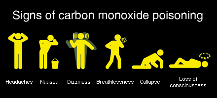
The clinical manifestations of CO poisoning can be subtle. Patients may be mistaken as having influenza (similar symptoms and both peak in winter months), presenting with headache, myalgias, confusion, tachypnea, tachycardia, presyncope and altered LOC. Classically, patients are described as having cherry red lips, though this is an insensitive sign. One retrospective study found the initial diagnosis to be listed as stroke, cardiac ischemia, encephalitis or seizure in multiple cases, highlighting the diagnostic challenge. Other acute symptoms include cardiac chest pain, and this has been described in 30% of cases. This is associated with increased mortality in CO poisoning. Chronic symptoms include the neuropsychiatric syndrome, which presents with personality changes, focal deficits, or cognitive impairment. This occurs with a month following poisoning and can persist for years. The mechanism is not clear, but may be related to myelin destruction from reactive oxygen species.

Because of the non-specific nature of CO poisoning a high index of suspicion is needed to make the diagnosis. The oxygen saturation on pulse oximetry is normal, given the maintained binding of hemoglobin. The diagnosis is made using co-oximetry to measure the amount of carboxyhemoglobin (venous or arterial blood). Amounts of carboxyhemoglobin can be found in several situations:

1-3% carboxyhemoglobin - Normal individuals
under 10% - smokers
Consider the patients history and presentation to determine if the levels are reasonable or as a result of poisoning.

The treatment of CO poisoning is emergency management.

1. ABC's - Many patients will have CO poisining from smoke inhalation. Security of the airway is crucial. Tachypnea, hypoventilation, stridor, facial blistering, burns and edema should prompt intubation and laryngoscopy. Patients can develop airway edema within 24 hours after the inhalation injury following thermal injury, this requires admission and monitoring for respiratory deterioration.

2. Oxygen therapy - all patients should receive high flow 100% oxygen. This results in rapid
improvement in CO levels compared to room air. The half life of CO is 300 minutes, and decreases to 90 minutes when supplimental oxygen is provided.

3. Consider hyperbaric oxygen therapy (HBOT) - Although this therapy is ubiquitous within the literature, the benefit of hyperbaric oxygen is somewhat controversial. There is no doubt that hyperbaric oxygen decreases the half life of oxygen even further than high FiO2 therapy, decreasing the half life to roughly 30 minutes. Whether this extends to improved long-term outcomes is debated. A cochrane review from 2011 found that further RCT's are required to determine whether HBOT should be offered, based on the poor quality of previous studies (see link below for details). Indications are variable, some directives suggests asymptomatic patients with a level of  over 40% CO should have HBOT, while others say 25% is a reasonable cutoff. Other reasons to initiate HBOT are:

Pregnancy and CO level over 20%
Decreased LOC or new neurologic deficits
Myocardial ischemia
Acidosis (less than pH 7.1)

4. Isocapneic hyperventilation - this is an experimental treatment that doesn't seem to be widely used in the clinical setting. One study (actually affiliated with anaesthesia at the University of Toronto), showed that in dogs, hyperventilating while providing inhaled CO2 to maintain pH balance resulted in improved removal of CO.

Cochrane review on HBOT


Wednesday, October 9, 2013

Rhabdomyolysis

Defined as dissolved skeletal muscle content, rhabdomyolysis is exactly that. It can present in many ways, from completely asymtpomatic to life-threatening end-organ damage.  In the early 1900's the syndrome of rhabdomyolysis was recognized as a cause of acute kidney injury following crush injuries. A case series corroborated this following the bombing of London in 1940, where patients injured from falling buildings were found to develop similar symptoms. Non-traumatic causes and the overall pathophysiology of rhabdomyolysis were not well recognized until decades later.


The final common pathway in causing rhabdomyolysis is increased intracellular calcium. Whether there is direct trauma to the sarcomere from trauma, or ischemia causing depleted ATP, all patients with rhabdomyolysis develop increased intracellular calcium. These leads to a worsening of intracellular energy stores, as Na/Ca exchangers actively use ATP to maintain normal electrolytes gradients. Increase reactive oxygen species leads to direct toxicity to cell membranes, and muscle death an intracellular content release occurs.

The causes of rhabdomyolysis are many, but can be broken down into several categories. I like the way its categorized on UptoDate, as it really allows you to structure your differential diagnosis. :

1. Traumatic - crush, trauma, immobolization from fall/coma (as in our patient today), burn, compartment syndrome

2. Non-traumatic exertional - significant exercise (marathon runner), myopathies that are exposed through exercise - (McCardle syndrome, carnitine palmitoyltransferasedeficiency)
Some people might include seizure in this category as it is a direct result of muscle contraction and exertion

3. Non-traumatic non-exertional - inflammatory myopathies, endocrine causes (hypothyroidism, DKA, pheo), infectious causes (viral, bacterial and parasitic causes - malaria), drugs (statin, alcohol, colchicine, herbal products, opioids, illicit drugs - cocaine) and electrolytes abnormalities (hypokalemia, hypophosphatemia)

The diagnosis can be suspected clinically, but we rely on laboratory findings to confirm rhabdomyolysis. The triad of weakness, dark urine and myalgias is typically present. On examination, muscles can appear swollen and tender on palpation. Additional urinalysis testing can be helpful. Myoglobinuria can be identified on dipstick at a level of 150mg/L, but can not be differentiated from hemoglobin. Microscopic examination will show minimal/no red blood cells if pure myoglobinuria. The pH of urine in this setting tends to be acidic. Proteinuria can be seen in up to 50% of cases. If there is more than 300mg/L of myoglobin in the urine, there will start to be a colour change to reddish/brown.

The main concern in managing rhabdomyolysis is the development of acute kidney injury. Its estimated that 10% of all AKI are related to rhabdomyolysis. However, the CK level doesn't correspond directly with increasing chance of AKI. That being said, renal injury usually develops at levels above 15,000. Mechanisms of renal injury include direct tubular toxicity, renal vscocrinstriction and tubule blockage due to myoglobinuria, not CK. This is interesting considering we measure CK levels, and not myoglobin levels in the serum (which are actually quite insensitive for the diagnosis if rhabdomyolysis). The direct tubular toxicity (mainly proximal tubule) is thought to be related to myoglobin biproducts, including free iron and production of reactive oxygen species, which can lead to direct tubular cell injury. Renal vasoconstriction is likely multi-factorial, given these patients are oven volume deplete, ill and reduced vasodilating agents (ie. NO). Tubular blockage occurs when myoglobin accumulates in distal tubules (most common). This process is enhanced by acidic urine, and volume depletion, which can cause increased Tamm-Horsfall protein casts with myoglobin can precipitate in.

Treatment is mainly focused on volume expansion to prevent AKI and treating the underlying cause. Certain IV fluids are promoted in the treatment for several reasons:

1. Normal saline - good volume expander if hypotensive/significantly dehydrated. Issues with this include the significant chloride load, which put patient at risk for metabolic acidosis. This may worsen some the physiologic mechanisms that can lead to myoglobin induced kidney injury (vasoconstriction, precipitation and tubular injury).
2. RL - reasonable choice given lack of chloride.
3. Sodium bicarbonate - will allow alkalinization of the urine, which will theoretically prevent the above issues with myoglobin. However, no RCTs have shown this strategy to improve outcomes. It seems reasonable to include this in therapy, especially if patients is hyperkalemic secondary to muscle injury.
4. Mannitol - suggested as a volume expander as it can lead to an osmotic diuresis and improved GFR, theoretically clearing tubules of myoglobin and cast debris.

Here is an article identifying some predictors for AKI in rhabdomyolysis.

Predictors of AKI and Rhabdo


Saturday, October 5, 2013

Cryoglobulinemia

Cryoglobulins are antibodies that precipitate at temperatures below 37 degrees Celsius. Its important to recognize that cryoglobulinemia is by definition the presence of precipitating proteins at low temperatures, however, clinicians tend to use this label to mean the clinical syndrome it is associated with. Cryoglbulins are classified into several categories, with each having its own clinical features and associations. :

Type 1 cryoglobulinemia - is monoclonal antibodies of one type, ie IgG or IgM. Associated with hematologic malignancies (Waldenstroms macroglobulinemia or myeloma). The primary clinical concern here is hyperviscosity syndrome where patients presents with infarcted digits, TIA/stroke like symptoms or thrombosis.

Type 2 cryoglobulinemia aka essential mixed cryoglobulinemia - results from monoclonal and polyclonal antibodies, including rheumatoid factor. The large majority of these patients have hepatitis C (30-100%), although connective tissue disease (Sjogren's syndrome) and lymphomas can also lead to this condition. Other less common infectious causes include HIV, streptococcal infections and brucellosis.

Type 3 cryoglobulinemia - is another form of mixed cryoglobulinemia, where all antibodies are polyclonal.

The classic triad was described by Meltzer in 1966, as purpura, arthritis and myalgia. These symptoms suggest cryoglobulinemic vasculitis. Palpable purpura is the most common manifestation, present in up to approximately 80% of patients. Purpura refers to bleeding under the skin that is larger than 3mm, when less than 3mm its termed petechiae. Other dermatologic findings include ulceration, levido reticularis and digital necrosis. Renal involvement is present in 30% of patients, where proteinuria, hematuria and active sediment production can occur. Nephrotic and nephritic syndrome occur in 21% and 14% of cases respectively. Neuropathy is also common, with up to 60% of patients describing symptoms burning and parasthesias. Mononeuritis multiplex (neuropathy involving a nerve with a name) is also seen, where patients can develop wrist or foot drop.

Testing for cryoglobulinemia is problematic, where samples need to be obtained in warmed syringes and kept at 37 degrees. Once serum is isolated it can be stored in a refrigerator at 4 degrees. It takes days for mixed immunoglobulin to precipitate, but type 1 proteins can be identified within hours. The quantity of cryoprecipitate can also be determined, using a test called the cryocrit. This is important because disease severity is proportional to amount of protein. Additional tests include complements, where low C4 is common, and elevated rheumatoid factor. Looking for an underlying disease association is also important if not already recognized.

Treatment is targeted towards the underlying cause and removing the monoclonal immunoglobulin. Considering HCV is so common in mixed cryoglobulinemia, ribavirin and interferon therapy should be considered with consultation by hepatology. Plasma exchange is helpful in life threatening disease with hyperviscosity. There is some growing evidence for the use of rituximab (monoclonal antibody targeting B-cells). A RCT from 2010 showed expedited rates of remission, improved renal involvement and higher rates of protein clearance compared to antiviral therapy in those with HCV. See a NEJM review on treatment in HCV associated cryoglobulinemia for more details (courtesy of the admitting physician).

HCV cryo treatment


Thursday, October 3, 2013

Heart block

Abnormal conduction between the atria and ventricles, termed AV conduction blocks, are common conditions in internal medicine. They range from mild asymptomatic findings to emergent, life threatening conditions. In 1964, Lev published the anatomic basis for atrioventricular blocks in the American Journal of Medicine, and still today, the disease that carries his name is responsible for nearly 50% of cases. The common nomenclature of AV block is documented as:

1st degree - slowed conduction between atria and ventricles with maintained synchrony
2nd degree - impaire conduction with intermittent loss of synchrony (separated into type I and II)
3rd degree - complete loss of synchrony between atria and ventricles. 

For the most part, conduction block are a disease of ageing or ischemia. Lev's disease, is used to describe the gradual fibrotic changes that occur with ageing to the cardiac conduction system, which lead to impaired functioning. Myocardial ischemia is the next most common cause, and can be acute or chronic. One study (Circulation, 1978) examined the natural history of heart block in patients following acute MI, and found that AV block is common. Twenty-two percent of patients developed second degree heart block type II. The most common type of blocks were LBBB and RBBB with anterior fascicular block. Patients who developed high grade AV block post MI had a significantly increased mortality (47% vs 23%), which was often directly related to the arrhythmia development and hemodynamic compromise. Other less common causes for AV block include:

Infection - endocarditis (aortic root abscess leading to AV pathway dysfunction), diptheria, scarlet fever, tuberculosis (with pericardial invovlement) mumps, lyme, toxoplasmosis
Inflammatory conditions - rheumatoid arthritis, lupus
Miscellaneous - sarcoidosis and amyloidosis (from infiltration)
Cardiomyopathy - hypertrophic cardiomyopathy, myocarditis'
Genetic - inherited forms of AV block exist, but are uncommon
Medications - remember to take a detailed history to identify agents that will impact the conduction system. 

Today, our patient was taking donepezil (an acetylcholinesterase inhibitor), which may have contributed to the development of bradycardia and heart block. There are multiple case reports of high grade AV block and even torsades des pointes with this class of medications. Donepezil is thought to be more associated with SA node dysfunction, causing sinus bradycardia more than high grade AV block, but both have been documented.  

After treating possible underlying conditions and removing offending medications, the next step is to consider pacing and pacemaker insertion.Determining treatment for the large majority of patients, which have ischemic or age related conduction disease, is based on symptoms and risk of worsening conduction delay.  
Generally accepted reasons for pacemaker insertion in AV block include:

1. Complete heart block, 
2. Symptomatic second degree heart block
3. Second degree block type II with consecutive dropped atrial contractions
4. Second degree block with previous wide complex on ECG
5. Exercise induced high grade blocks

Patients with bifascicular and trifascicular block may also require a pacemaker in the setting of syncope, given it may be due to the development of transient third degree heart block. More detailed instructions can be found in the AHA guidelines for pacemaker insertion linked below.


Wednesday, October 2, 2013

Enterococcus and endocarditis

Infective endocarditis (IE) is a huge topic. The IDSA guidelines are very comprehensive and cover the treatment of IE based on microbiology and valve involved (mechanical vs bioprosthetic). These are linked below.

Enterococcus fecaelis and fecium were given their name to emphasize their presence in the human gastrointestinal tract. There are multiple species of enterococcus, but these two tend to be the most clinically relevant bugs. In the 1930's, the Lancefield classification placed enterococcus in group D, along with strep gallolyticum. Enterococcus was identified as a cause for IE as ealy as 1906, and over the following decades found to cause urinary tract infections, biliary infections, peurperal sepsis, and wound infections during WWI. 

Infective endocarditis is caused by enterococcus in 5-15% of cases. The virulence of this organism is much lower compared to other organisms (ie. S. aureus). Two studies from the 1980's found that bacteremia from enterococcus had a relatively low likelihood of developing endocarditis, where only 2.5% of those with positive blood cultures met criteria. E. fecaelis is more common than E. fecium, which often requires vancomycin therapy given penicillin resistance. Even less frequent than this is vancomycin resistant enterococcus as a cause for endocarditis.  Enterococcal endocarditis is more common in men and older patients, where the average age in one study was 65.  One study from the archives on internal medicine showed that 50% of cases in men followed urinary tract instrumentation, an important risk factor that was present in our case discussion. Other associations include a history of malignancy and nosocomial acquisition. Below is a relatively recent retroprospective database from the American Jounral of Medicine.

Anecdotally, enterococcal endocarditis is less commonly associated with peripheral stigmata, which were absent in our patient. The disease course tends to be subacute with aortic valve involvement being most common.  

Unfortunately, enterococci are less susceptible to beta-lactam antibiotics and synergy with aminoglycosides are often employed. The purpose of dual therapy is to have cell wall breakdown from the penicillin antibiotic allow for improved penetration of the aminoglycosides, which act intra-cellularly on ribosomal activity. Different strains can have varying susceptibility to these antibiotics, which may influence dosing and antibiotics choice. Gentamicin/streptomycin have toxic effects, including ototoxicity and nephrotoxocity and the minimum six weeks of antibiotics required for this diagnosis can lead to significant morbidity. Prosthetic valve endocarditis may fail to be effectively treated with antimicrobials alone, and often requires surgical opinion. I would also advocate for the involvement of the ID service to help with antibiotic choice, dose, interval and duration of therapy.

Tuesday, October 1, 2013

Chronic myeloid disorders

Chronic myeloid disorders can be a confusing group of diseases to understand. There is overlap between conditions, making diagnosis challenging at times and many conditions have multiple names. Today we discussed a case of myelodysplastic syndrome, which is very likely a case of myelofibrosis. Here, we describe a general approach to these conditions and some of the clinical features.

Chronic myeloid disorders is an umbrella term encompassing multiple conditions: chronic myeloid leukemia (CML), myelodysplastic syndrome (MDS), chronic myeloproliferative disease and atypical chronic myeloid disorders. Chronic myeloproliferative disorders can be broken into polycythemia vera (PRV), essential thrombocytosis (ET) and idiopathic myelofibrosis (IMF). I think that confusion arises as a result of the multiple subgroups within each category of disease. You need to read about each entity separately to gain a better understanding of its differences.

MDS is as a result of ineffective and disordered hematopoeisis in one or more cell lines. Despite low peripheral counts, bone marrow evaluation reveals normal or hypercellularity. This is generally a disease of the elderly, where the majority of patients are over 65. It can be seen earlier, increasingly if caused by an external chemical exposure (ex. benzene). Clinical features are those associated with low blood counts; infection, bruising, fatigue etc. Some additional hints towards a diagnosis include an increased number of band neutrophils on the smear, the so called "pseudo-Pelger-Huet" anomaly, a result of premature cells being pushed into the blood.

The classification of myelodysplastic syndromes (MDS) changed in 2001. The World Health Organization changed the previous French-American-British (FAB) definition to improve the diagnosis of AML. MDS are a group of myeloid neoplasms, that impact on the proliferation of hematopoietic cells. Patients can present in a myriad of ways, and are generally classified into multiple categories:

1. Refractory cytopenia with unilineage dyslpasia- low RBCs or low platelets or low neutrophils
2. Refractory cytopenia with multilineage dysplasia- multiple low cell lines
3. Anemia with ringed sideroblasts- sideroblasts are abnormal mitochondria that are visibly different as a result of increased iron content
4. Refractory anemia with excess blasts - two stages based on percentage blasts (5-10%, 10-20%)
5. MDS from 5q deletion- a specific genetic variant
6. MDS otherwise unclassified

Prognosis varies depending on the subtype of MDS. Twenty percent of patients with MDS will die from AML, however those with 5q deletion are less likely to convert, and have a more indolent course.

Myelofibrosis (IMF), aka agnogenic myeloid metaplasia, is a different disease. This is a clonal stem cell disorder with progressive bone marrow fibrosis. An abnormal precursor is thought to produce factors that result in fibroblast proliferation and collagen deposition. Premature fibrosis pushes precursor cells into the periphery resulting in deposition of these cells in distant tissues. This leads to hematopoeisis outside the marrow. Cells can deposit in many different organs, including the lungs, pericardium, spleen, bowel, liver etc. IMF is less common than MDS, present in only 0.5 people per 100,000. The clinical presentation includes fatigue, splenomegaly, constitutional symptoms, and complications of bone marrow failure. Counts are often increased initially because of compensatory extramedullary hematopoeisis. Blood smear will often show premature WBCs, teardrop cells, giant platelets, termed a leukoerythroblastic pattern. Up to 60% of these patients will have a testable mutation in a gene called JAK-2. Occasionally,  can be difficult to distinguish IMF, from burnt out ET or PRV.

The prognosis in IMF is poor, with 10% of patients transforming to AML, having an average survival of 2-5 years. Unfortunately there are few successful treatments for this condition. In young individuals, allogeneic stem cell transplant can be considered, but in the elderly there is little that can be done. Blood counts can be controlled with various chemotherapy, and low counts managed with transfusion.

Two reviews for each of these conditions are listed below.

MDS review - NEJM
Myelofibrosis and NEJM


Thursday, September 26, 2013

HCC

Hepatocellular carcinoma (HCC) is a growing concern. Currently, it is listed in the top ten most common cancers worldwide amongst both men and women, and is continuing to increase. The majority of cases are in the setting of viral hepatitis, where hepatitis B makes up nearly 50%  of cases. Any causes of cirrhosis can theoretically lead to HCC, and rarely, HCC can develop in the absence of cirrhosis (10%). The burden of hepatitis C has been driving HCV in North America, where HCV related liver cancer is the fastest growing cause of cancer related death in the US.

Patients with HCC tend to present with symptoms of cirrhosis. Jaundice, ascites, GI bleeding and so forth. HCC should be considered in those presenting with acutely decompensated cirrhosis, as it can precipitate worsening liver disease through tumor extension. As an internist, it's also interesting to recognize the various paraneoplastic symptoms of HCC; erythrocytosis, hypoglycemia, sign of leser trelat, hypercalcemia, and diarrhea are recognized phenomenon. 

The diagnosis is based on imaging and possible need for biopsy. MRI or CT can be very helpful and when patients have predisposing conditions (HBV), a tumour of 2 cm or greater with typical radiographer pattern in pathognemonic for HCC. Early arterial enhancement and delayed venous washout on a multiphasic scan are suggestive, and indicate tumor vascularity. Tumours less than 1cm. Are difficult to biopsy and require serial scans for monitoring. Intermediate size nodules with atypical pattern should be followed with consideration of biopsy for definitive diagnosis. There are guidelines available on how to approach these nodules which can be very helpful. Alpha-fetoprotein (AFP), when over 500 mcg/L is diagnostic for HCC but intermediate levels can be seen in cirrhosis alone and results need to be taken in context. Levels over 200 mcg/L in the appropriate context have a specificity of over 95%.

Monitoring for HCC is suggested to include liver ultrasound and AFP every 6-12 months in those at risk. This is based on a study published in 2004, where nearly nearly 19,000 Chinese patients with HBV underwent this surveillance. They found a reduction on mortality of 37%. However, A study performed the year prior found no benefit. That being said, this is recommended by the current guidelines. patients that should be monitored include: 

1. Asians men over 40 with HBV
2. Asian women over 50 with HBV
3. Patients with HBV and cirrhosis
4. Blacks with HBV 
5. Family history of HCC and HBV
6. Any patient with cirrhosis

It is less clear when to start screening Caucasians with HBV, and some recommend initating at similar times to the Asian population, despite a lower risk of developing HCC. It may less cost effective to screen in those with cirrhosis from alcohol given a lower risk of HCC, but the epidemiology is not well described. Attached below is a nice NEJM review which contains many of the resources with above information.

HCC review


Wednesday, September 25, 2013

Hypoglycemia

Hypoglycemia is dangerous and common event for many patients with diabetes. Although it is more common in DM1, many patients with DM2 will experience this problem. The Diabetes Control and Complications Trial (DCCT) identified hypoglycemia as a major concern, causing symptoms approximately 2 times a week in those with DM1. As patients live longer with DM2, they also develop an increased frequency of hypoglycemia, where 25% of patients using insulin for over 5 years experience this problem regularly. Much of our focus when caring for patients with diabetes is lowering their blood sugar in the hopes of preventing complications. Microvascular complications are reduced by improving glycemic control in both patients with DM1 and DM2, making it the focus of diabetes care. However, randomized trials targeting lower HbA1c levels (ex. ACCORD), found increased mortality in those randomized to tighter glucose control. Although not directly proven, this is likely a result of increased hypoglycemia. This highlights that severe symptoms can occur as a result of hypoglycemia. A study published in Diabetes Care found 100% of patients with DM1 experienced severe hypoglycemia at some point, requiring medical intervention. Recognizing the problem is an opportunity for prevention. Many diabetics with recurrent episodes lose the ability to sense hypoglycemia, putting them at risk for lower levels and additional risks. Symptoms can be broken down into two categories:


1. Neurogenic symptoms - tremor, palpitations, arousal, sweating, hunger (mediated through catecholamine and acetylcholine mechanisms).
2. Neuroglycopenic symptoms - cognitive impairment, coma, seizure and death.

Patient who experience recurrent hypoglycemia are at increased risk for developing hypoglycemia unawareness. This is when, despite hypoglycemia, patients lack symptoms to suggest a problem. Often the first symptom in these patients is confusion, which makes it hard for them to respond appropriately with increased sugar intake. Hypoglycemia unawareness can lead to hypoglycemia associated autonomic failure (HAAF), a form of autonomic insufficiency from recurrent hypoglycemia. In HAAF, patients are unable to mount an appropriate adrenergic response to low blood glucose. HAAF is associated with a significant risk of severe hypoglycemia (25x those without). If glucose levels improve and are maintained, patients will regain the function of their catecholamine axis in several weeks.

Definitions of hypoglycemia are different whether or not a diagnosis of diabetes is present.In those with diabetes any glucose less than 3.9 mmol is considered low. In those without diabetes the guidelines state that patients must have whipples triad:

1. Symptoms that may be explained by hypoglycemia
2. Documented hypoglycemia at the time of symptoms
3.Improvement of symptoms after taking glucose

There are many causes of hypoglycemia, and the approach is different in those with and without diabetes. The most common cause of low glucose is druge related. Several categories exist and can be separated by either "sick patients/medicated" or "well patients":

"Sick/medicated patient"
1. Drugs-insulin, alcohol
2. Critical illness- sepsis
3. Organ failure- renal/liver failure
4. Cortisol deficiency
5. Non-islet cell tumours
"Well patient"
1. Endogenous overproduction of insulin- insulinoma, nesidioblastosis
2. Autimmune- antibodies against endogenous insulin
3. Use of secretagogue- accidental, serruptitious etc.

More details regarding investigations for hypoglycemia and management of this illness in patients with diabetes can be found in the guidelines linked below.

Hypoglycemia guidelines


Thursday, September 19, 2013

Suspected overdose

Paracelsus, the Renaissance physician said that "the dose determines that a thing is not a poison", recognizing that most things in excess can be harmful. In the US there are over 2.4 million documented toxin exposures reported to poison control centres, the majority of which occur in children. The leading agents causing death are analgesics, antidepressants, cardiovascular medications, stimulants and illicit drugs.

Your approach to the poisoned patient is two pronged, containing a diagnosis and a treatment are in parallel. The treatment consists of ABC's, D (decontamination/DONT antidotes), E (enhanced elimination), F focused therapy and G (get help). The diagnostic are requires a history, physical exam, search for toxidrome and diagnostic testing.

History is key. How much? When? What? Why? All these can be helpful and alter your approach to the patient. Often patients cant name the medications they're taking or they do so incorrectly (Tylenol vs aspirin). If it is not available, obtaining information from family, work, family physician may be helpful. Specific symptoms to consider include:

protracted coughing - hydrocarbon exposure
inability to swallow/drooling - costic ingestion
hematemesis with iron ingestion
persistent seizures with INH overdose
decreased LOC with carbon monoxide

There are a number of pneumonics to help you remember toxidromes. Below is an article that contains an extensive list. Things to focus on include vital signs (HR, BP, temp, GCS, seizure), pupils (meiosis, mydriasis), odour (garlic-organophosphate, wintergreen - methylsalicylates, almond - cyanide), neurologic exam (rigidity/clonus - SS/NMS), skin (rash, diaphoresis, track markrs, fentanyl patches).

Preliminary labs will include routines, plus toxicology screen, osmolal gap and anion gap. Toxicology can be performed on serum and urine, where urine test tend to identify metabolites and will have positive results for longer duration compared to serum. Quantitative levels should only be performed if it suggests higher toxicity and will alter management. Levels can be performed on: tylenol, ASA, Li, Fe, CO, digoxin, anticonvulsants, toxic alcohols and theophylline. Other aspects of urine testing include any visual change, such as a change in UV light with ethylene glycol, orange with rifampin, pink with ampicillin, green with copper or methylene blue. Microscopy for calcium oxalate crystals also suggests ethylene glycol poisoning (see link below). Imaging tests may include CXR/AXR to look for iron or ingested street drugs (body packing with cocaine). Drugs causing pneumonitis or pulmonary edema can be remembered as MOPS (methadone, opioids, phenobarbitol/phosgene, salicylates).

Today we mentioned that "one pill can kill", although this tends to be targeted more towards children it highlights the potential toxicity of these medications. A top ten list of these drugs includes, but is not limited to:
1. TCA
2. antipsychotics
3. antimalarials
4. anti-arrhythmics
5. camphor
6. oral hypoglycemics - sulfonylureas
7. opioids
8. theophylline/podophylline
9. salicylates
10. calcium channel blockers

Calling poison control is always the right thing to do. You will be put in touch with an experienced nurse and have access to a clinical toxicologist if necessary. They also document each case and follow-up on the patient. For a more detailed approach to therapy see the article below.

Approach to unknown overdose
calcium oxalate cyrstals
fluorescent urine in ethylene glycol ingestion

Wednesday, September 18, 2013

Alcohol use - keeping an eye open

The complications of alcohol use are many. We are very good at listing the extensive number of possible problems that come along with alcohol use; nutritional deficiencies, myopathy, cognitive impairment, cirrhosis, cardiomyopathy, etc. however we are often fail to characterize the problem. Physicians underestimate the amount of alcohol ingested and when its identified, fail to capitalize on opportunities for rehabilitation. A US study found that only 48% of patients identified as having problem drinking were asked to follow-up, leaving the majority without ongoing monitoring. Patients may fail to have recognizable signs of cirrhosis despite significant liver injury, requiring probing questions to first identify the problem.


There are many tools for evaluating alcohol abuse. The most commonly used tool is the CAGE questionnaire. This method is great because of its simplicity and ease of administration, but fails to determine long term or changes in drinking patterns.It also lacks a quantitative component, which can be helpful to gauge severity of disease. A meta-analysis evaluating its use found the test to be more useful in inpatients compared to ambulatory assessment with a sensitivity of 87% and specificity of 90% for >2 components. Other tests do exist.

The alcohol use disorders identification test (AUDIT) is a longer test with higher sensitivity and specificity (96% and 97%). This was developed by the World Health Organization, and as a result was created for international use, validated in multiple languages. Of its 10 questions, it covers consumption, consequences and dependence issues, covering more areas than shorted questionnaires. Values of 8 and 20 are important to remember, being markers of harmful alcohol use and dependence.

Other markers of alcohol use which are mentioned on the wards and in the literature are macrocytosis and GGT levels. Unfortunately, these tests have limited sensitivity and specificity. A study in Hepatology from 1984 found GGT did offer some prognostic information in patients with acoholic cirrhosis, where only 60% of those with a level of 100 IU survived to 1 year.

An additionally proposed test for alcohol consumption is measurement of carbohydrate deficient transferrin. Transferrin circulates in our bodies in glycosylated forms. There is a variety of sugars attached to transferrin in different amounts. Alcohol consumption results in reduction of carbohydrate numbers on the transferrin molecule, something that can be detected in the blood. Studies have found that patients with low AUDIT scores are unlikely to have carbohydrate deficient transferrin. The test characteristics have been proposed to be better than that of GGT. I am not familiar with the cost of this assay and have never heard of anyone ordering this test.

All patients admitted with alcohol related complications, and more importantly those seen as outpatients with risk factors for alcohol abuse should be screened. Multiple methods exist, but when you have time the AUDIT form seems like an appropriate choice. See additional details below.

AUDIT score from BMJ best practices

Friday, September 13, 2013

Malignancy and diarrhea

Infectious causes dominate the majority of causes for acute diarrhea, and its often what we first consider based on investigations, and possible treatments. However, with diarrhea lasting more than 2 weeks, persistent and chronic diarrhea are considered, and after the infectious group we have to think about other classes of disease.

Malignancy is an uncommon but ominous cause of diarrhea. This association can result from several ways, and categorizing the type of diarrhea can be helpful. Typical groups include the following:

1. Watery diarrhea - secetory vs. osmotic
2. Fatty diarrhea
3. Inflammatory diarrhea

Watery diarrhea can be identified on history, where patients tend to have large volume, frequent stools, without solid material. Patients will often describe diarrhea despite not eating, and having to wake in the night to have a bowel movement in secretory/osmotic causes. Although its often not necessary, the stool osmolality gap can be calculated to differentiate between osmotic and secretory where a gap of greater than 125 suggests a osmotic cause. Cancers causing secretory diarrhea include the islet cell tumours which secrete hormones including: gastrinoma, glucagonoma, vasoactive intestinal peptides tumours, and pancreatic polypeptide tumours. Increased seratonin is also felt to be the cause of secretory diarrhea that occurs in carcinoid syndrome. Patients with medullary thyroid cancer complain of diarrhea as a ommon symptom in metastatic disease present in 40% of patients. The mechanism is controversial, but calcitonin production is thought to play a role. The diarrhea in this disease was clasically considered a secretory diarrhea, though some studies have found an increased electrolyte gap in the stool suggesting an osmotic cause. Lymphoma (when present in the gut) and villous adenomas of the bowel are also felt to produce a secretory pattern of diarrhea.

Fatty diarrhea is diagnosed using a 72h quantitative fecal fat test. Additional sudan staining can confirm the presence of fat in the stool. Patients will often say that they have greasy, oily stool that doesnt go down the toilet after flushing because it floats. Malabsorption from multiple causes can present like this, the most common likely being celiac disease. However, again malignancy can cause a similar presentation. Pancreatic cancer that results in exocrine dysfuntion can impair fat digestion in the bowel and lead to fatty diarrhea. Biliary obstruction from pancreatic cancer will decrease the release of bile salts in to the bowels and again impair fat absorption as a result, contributing to diarrhea. Somatostatin secreting tumours are documented as causing fatty diarrhea, these are islet cell tumours that produce octreotide, inhibit pancreatic function and bicarbonate release, which results in fat malabsorption. 

Many patients with cancer are on chemotherapy and are therefore immunosuppressed. Diarrhea may be a direct result of chemotherapeutic agents, or newly acquired infection. Our first focus of care in diarrhea should be to rule out infection and avoid exacerbating medications given these things can be life threatening and are treatable. 

Thursday, September 12, 2013

Renal artery stenosis

In hospitalized patients acute kidney injury is largely attributed to pre-renal disease, with additional renal and post renal causes being much less likely. Although a trial of fluids is usually tried in the majority of patients its important to recognize when this therapy is failing and other causes of renal should be considered. Renal artery stenosis (RAS) is an uncommon cause of chronic hypertension (less than 1%), but is more likely to be causative in acute refractory cases. In general, RAS can be broken into two classes:

1. Atherosclerotic stenosis (90%)
2. Fibromuscular dysplasia (10%)

Atherosclerotic disease is much more common than FMD given the presence of CAD, DMII, HTN, dyslipidemia and smoking prevalence. It is a progressive disease which usually results in small kidneys and CKD. Fibromuscular dysplasia is an abnormality of the intimal, medial and adventitial layers of the blood vessels of unclear cause. It results in narrowing of the renal arteries, typically at the distal third. Risk factors include being female and HTN prior to age 50. These patients can develop other complications, including artery dissection or thrombosis (most common with intimal involvement).

The pathophysiology is related to poor renal perfusion leading to activation of the renin-angiotensin system, impacting sodium homeostasis, vasodilatory factors and results in renal hypertension. Clinical clues to suspect renal vascular disease in patients with CKD and HTN include:

1. Worsening kidney function of 30% in started and ACEi
2. Systolic/Diastolic renal bruit
3. Onset HTN after 55
4. Flash pulmonary edema
5. Asymmetry in renal size >1.5 cm

Testing for these disease used to involve a captopril renal scan where differences in renal perfusion were examined following administration of an ACEi. However, this is no longer done and has largely been replaced by imaging. Ultrasound with arterial dopplers are helpful, and certain flow velocities through the artery can predict the degree of stenosis. This does however require an experienced radiologists and can be difficult to get appropriate view. CT angiography and MRI are useful tests, but are less likely to image distal vessels appropriately.

All patients are recommended to undergo medical therapy, which involves modification of CAD risk factors, including smoking cessation. ACEi will preferentially vasodilate the efferent renal artery, counteracting the natural response to improving GFR in patients with afferent renovascular disease. The addition of an ACEi can decrease GFR and worsen AKI. That being said, they can still be used in this disease and one must consider the clinical context. This is more problematic inpatients with atherosclerotic disease as opposed to FMD.

Renal artery angioplasty has not been shown to be superior to medical therapy in a meta-analysis published in the American Heart Journal in 2011. However, patients with refractory hypertension and flash pulmonary edema, may be considered for angioplasty. See this NEJM review on RAS for additional details.

Renal artery stenosis review

Wednesday, September 11, 2013

Thiamine and lactic acidosis

The approach to a lactic acidosis involves separating it into two possible classes, termed type A or type B. Type A is classified as lactic acidosis secondary to tissue hypoperfusion. This is usually not a mystery, and the patient has signs of end-organ damage, tissue hypoxia and hypotension. Type B lactic acidosis is felt to be from some derangement in metabolism of lactate, including:


1. Drugs - metformin use, HIV medications (through mitochondrial dysfunction)

2. Alcohol - decrease gluconeogenesis

3. Malignancy - suspected to be secondary to increased cell turnover, though the mechanism is somewhat unclear, this is more often seen in leukemia/lymphoma

4. Type-D-lactate- present in patients with short gut syndrome, bowel bacteria (usually lactobacillis) produce D-lactate which is absorbed but cannot be metabolised by endogenous LDH leading to accumulation.

5. Liver dysfunction

Today we discussed a patient that had an elevated lactate of unclear cause. The patient was treated with thiamine with rapid improvement, which raises nutritional deficiency as a potential contributor to type B lactic acidosis.

Thiamine (vitamin B1) is a water soluble molecule that is required for aerobic metabolism. It acts as a cofactor for several enzymes present in the glycolysis pathway. Thiamine deficiency prevents pyruvate from converting into Acetyl-CoA, which is the entry point into the Krebs cycle, which is responsible for ATP production and NADH which is utilized in the electron transport chain. As a result, pyruvate accumulates and is metabolized into lactate resulting in acidosis.

Thiamine deficiency can occur quickly. Because it is water soluble it is not readily stored in fat and levels lowering the total body amounts. It is estimated that thiamine deficiency can develop as quickly as four weeks in poor nutritional intake. In cases of thiamine deficiency lactate production has been seen as early as 1-3 weeks. Risk factors for thiamine deficicency include, poor nutritional intake, folate deficiency, alcohol use, malabsorption, TPN and renal disease (specifically dialysis/peritoneal dialysis).

So next time you see a lactic acidosis in the absence of tissue hypoperfusion, consider vitamin B1 deficiency.

Tuesday, September 10, 2013

VBG versus ABG

The purpose of performing an arterial blood gas (ABG) is to identify alterations in acid base status or ventilation/oxygenation. Arterial blood gases can be troublesome at times. Patients may have hemodynamic instability (miking it tough to find the pulse), be unable to hold still for sample collection, and the procedure can occasionally be technically difficult. As a result, venous blood gases are often taken as a surrogate for arterial measures given they are easier to perform, less painful, and patients in the ICU often have central venous access.


There are several different areas from which a blood gas can be drawn:

1. Arterial blood gas - usually radial artery, though can be taken from brachial/femoral stab
2. Peripheral venous blood gas - any peripheral vein
3. Central venous gas - from right atrium, usually drawn from central line
4. Mixed venous gas - taken from the distal port of pulmonary arterial catheter

Of these different types of blood gas', the correlation with ABG's are variable:

1. Central venous vs ABG - pH 0.003-0.005 lower, pCO2 4-5 mmHg higher, no change in HCO3
2. Mixed venous vs ABG - similar to above
3. Peripheral venous vs ABG - pCO2 3-8 mmHg higher, HCO3 1-2 meq higher

pO2 values can not be reliably calculated from any form of venous gas.

Of these options central venous gas is preferred given its correlation with the ABG is most consistent and most studied, that being said, many patients dont have central lines.There was a study examining the effects of tourniquets on venous blood gas values and found that their use doesnt alter blood gas variables when used, which is reassuring.

The clinical context needs to be considered. If pateints are hypotensive and in shock, venous values are less reliable and ABG's are preferred. A study from NEJM in 1989 showed that as patients become more unstable, the arterial-venous difference gets larger. The difference between central venous and ABG pH in patients with shock was 0.1, highlighting that both measures need to be evaluated. They also concluded that tissue hypoperfusion is better assessed with central venous gas than ABG. Another NEJM study looked at a similar question in patients during cardiac arrest. The average ABG pH with during CPR was 7.41, while the mixed venous was 7.15! The pCO2 also changed dramatically. ABG may not reveal the true extent of tissue hypoxia compared to the mixed or central venous blood gas.

So, when in doubt, I say get an ABG, but in the critically ill patient/post CPR, a venous gas may show the true extent of the disease.


Thursday, September 5, 2013

UTI prophylaxis

Recurrent urinary tract infections (UTI), defined as 2 or more infections in six months, or over three in a years period, is a frustrating disease for patients and physicians. This disease is common, where 20% of women presenting with a UTI will experience a recurrent episode at sometime. For the most part, these are new infections as opposed to persistence of the previous infection. When encountered, it raises the question as to whether patients should be treated with antibiotics on a more frequent basis in a prophylactic manner. Many women experience symptoms in relation to sexual activity, though in post-menopausal women, recurrent UTIs may have a different mechanism. Ageing results in changes in bladder function and anatomy, with increasing risk of prolapse. This can lead to urinary stasis and predisposition to recurrent UTIs. There are in general three approaches to this topic:


1. Continuous antibiotic prophylaxis
2. Post-coital prophylaxis
3. Intermittent therapy (not really preventative)

Choice of prophylactic strategy should be tailored to the individual patient. Cultures should be taken from the urine to confirm the presence of recurrent infection. Antibiotic choice should be based on culture and sensitivity results.

For women with recurrent UTI's, not in relation to coitus, continuous prophylaxis can be prescribed to patient as daily or three times weekly regiments. Usual antibiotics include: septra, nitrofurantoin, cephalexin, ciprofloxacin. Several studies have shown decreased rates of infection with this approach, and in 2004 a Cochrane review was published finding similar results. The number needed to treat in this review was 1.8 for one years therapy (which was the duration in the majority of studies). There was however increased side-effects in the therapy groups compared to placebo, where GI upset, rash and vaginal irritation was most common. In this study, there was no supreme antibiotics that performed better compared to others.
Following discontinuation of antibiotic therapy, women will tend to revert back to having recurrent UTI's. A reasonable approach is to trial antibiotics for 6 months, and after stopping see if the patient improves. Some individuals will have clusters of infection, which will be best treated with a period of drug followed by a holiday period. Longer prophylactic periods, upwards of two years, have also been advocated by some in the literature.

For females that develop infections in relation to sexual activity a post coital strategy can be taken, and has been found to have similar rates of success to continuous prophylaxis.  One study randomized 135 women to daily prophylaxis with 125mg of cipro versus a post coital strategy. They found similar results between groups and a third of the amount of antibiotic used. A study published in JAMA 1990, comparing placebo to septra using a post-coital strategy showed a significant reduction in UTI's. Where 81% in the placebo group developed a recurrent UTI compared to 12.5% in the therapy arm.

Intermittent therapy that is self administered is a useful approach for patients who are compliant and motivated. This strategy results in a higher number of infections, given it is not a truly preventative approach, but symptomatic duration and total antibiotic dosing is minimized. One study testing this approach found that symptomatic episodes had a culture negative rate of 14%, suggesting that women were able to accurate identify the presence of a UTI based on symptoms in over 85% of cases.

With the ever-growing use of antibiotics, resistance to these medications is a concern, and should be considered in patients receiving chronic therapy. There have been studies of patients taking chronic antimicrobial therapy for UTI prophylaxis identifying breakthrough infections with bugs that were resistant to the antibiotic prescribed. Separate studies using septra and cipro found breakthrough infection rates with resistant organisms at a rate of 44% and 3% respectively. This high rate of septra resistant organisms in this group is concerning, and provides some evidence that its use as prophylaxis results in significant alterations in microbe susceptibility patterns. See this NEJM review from 1993.

Urinary tract infections NEJM