Monday, September 28, 2009

Clubbing


Today's physical exam rounds were on clubbing. Some points:

Etiologies (not meant to be exhaustive!)

1) Pulmonary- neoplastic, suppurative (i.e. abscess, empyema), bronchiectasis, inflammatory (e.g. fibrosis), pulmonary AVMs (as in HHT)
2) Cardiac- cyanotic heart disease, endocarditis
3) GI- cirrhosis (often non-alcohol related, as in PBC), celiac, IBD
Others: alpha-1 antitrypsin deficiency, sarcoma, hyperthyroidism


Classically first appears on 4th finger on non-dominant hand

Physical examination for clubbing:

May see obvious changes of terminal phalanges

Nailfold angles
1) Profile angle (AKA Lovibond's angle): Between skin proximal to cuticle and "takeoff" of nail; should be less than 180 degrees
2) Hyponychial angle: between skin proximal to cuticle and distal nail; should be less than 192 degrees.


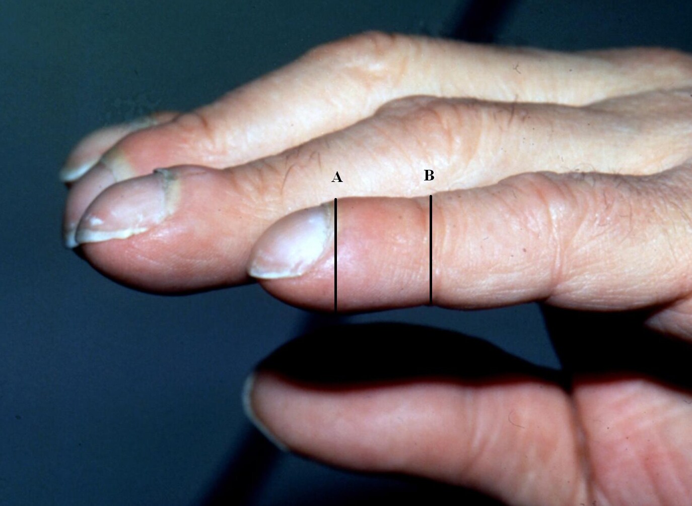
Phalangeal depth ratio
Ratio of the distal phalangeal depth (i.e. vertical height of the finger at the middle of the distal phanynx) to the interphalangeal depth (i.e. vertical height of the finger at the DIP joint). Interphanangeal depth is normally greater than distal phanangeal depth. This is reversed in clubbing.

Schamroth sign
Diamond-shaped opening when dorsal distal phalanges are opposed

Palpation:
May have "boggy" nailbed, where proximal edge of nail is felt, and may be able to rock the nail back and forth using nailbed as a fulcrum

Evidence:
Based on the JAMA Rational Clinical Exam article on clubbing,
the lack of an accepted gold standard makes sensitivity and specificty determinations difficult. Their literature review showed that 'normals' are very unlikely to have profile angle over 176 degrees, hyponychial angle over 192 degrees, or phalangeal depth ratio over 1.0. Patients are more likely to have IBD or lung cancer if clubbing is present, and the phalangeal depth ratio was the best test for this purpose.


Tuesday, September 22, 2009

The poisoned patient


Today we discussed an approach to the poisoned patient. Some points:

Detective work is often required in these situations, and history from witnesses / bystanders / family members / paramedics, etc. often provides a key clue

Always ask about

1) Details of what was found near the patient and events (including number of pills left in bottles- gives the "worst case scenario" in terms of amount of a substance taken)

2) Timing of ingestion

3) History of what medications the patient was taking, including possibly from pharmacy directly
4) Medications/substances the patient would have had access to (including family members' medications)

5) History of previous intoxications, psychiatric history

In many cases, collateral history is not possible, and even the most thorough searching does not reveal the substance taken. In these cases, the physical exam often reveals a "toxidrome" that can point to the substance and guide therapy.

Toxidromes (and some mnemonics)

Sympathomimetic
febrile, flushed, tachycardic, hypertensive (more than anticholinergic), mydriasis, diaphoretic (distinction from above)
Examples: cocaine, amphetamines, pseudoephedrine
Treatment: Benzodiazepines, supportive

Narcotic
dec. LOC, hypotension, dec. respiratory rate, miosis. Response to naloxone
Examples: Morphine, other opiates
Treatment: Naloxone

Anticholinergic
hot as a hare, dry as a bone, red as a beet, mad as a hatter, blind as a bat febrile, flushed, tachy, mydriasis, dry mucosa, decreased bowel sounds, urinary retention, hallucinations Examples: Benadryl (or other antihistamines), Gravol, tricyclic antidepressants, antiparkinsonians
Treatment: Acetylcholinesterase inhibitor (e.g. physostigmine)

Cholinergic:
"SLUDGE and the killer B's": salivation, lacrimation, urination, diaphoresis, gi (diarrhea), emesis. Killers: bradycardia, bronchorrhea
Examples: Organophosphate insecticides, donepezil, rivastigmine, galantamine
Treatment: Atropine

Sedative / hypnotic:
dec. LOC, dec. respiratory rate, slurred speech, ataxia, nystagmus
No response to naloxone
Examples: Phenobarbital, alcohols, benzodiazepines
Treament: No specific antidote for phenobarbital; flumazenil for benzo; EtOH or fomepizole for MeOH


Managing the poisoned patient:

1) ABC- may need definitive airway management, supportive care

2) Try to identify agent or toxidrome as above
Always remember co-ingestions! Ways to identify these are by the anion gap, osmolal gap, ECG (looking for signs of cardiac toxicity), and serum + urine toxicology screens

3) Administer "universal antidotes" where appropriate- oxygen, glucose, naloxone, thiamine

4) Think about preventing absorption
Activated charcoal or whole bowel irrigation
Charcoal: If less than 4h from ingestion (preferably less than 2h). Want 10:1 ratio of charcoal to substance. Not for caustics, metals (Li, Fe), hydrocarbons. Repeated charcoal with drugs with enterohepatic circulation- theophylline, phenytoin, carbamazepine
Whole bowel irrigation: Enteral PEG-LYTE administration until rectal effluent is clear. It is effective with drug packets, extended release preparations, substances not well adsorbed by activated charcoal. Dose 2L per hour PO or NG until clear.

5) Think about a specific antidote if one exists

6) Think about enhancing elimination
(e.g. by dialysis or alkalinization of urine)

7) Always call poison control!

Dialyzable drugs:
Salicylates, alcohols, Li, phenobarb, valproate, theophylline, carbamazepine

Links:

Click here for a NEJM clinical case outlining approach to the poisoned patient

Click here for a review of a new potential antidote to lipophilic drug intoxicatios, a lipid emulsion- "Intralipid"

Monday, September 21, 2009

Aortic insufficiency


Today's physical exam rounds were on aortic insufficiency. Some key points:


May be caused by disease involving aortic leaflets or root.

Valvular: Calcific (some element of AI in 75% or pts with AS); endocarditis, trauma (ascending aortic tear), rheumatic disease.
Root: Age-related, cystic medial necrosis (isolated or with Marfan's), bicuspid valve, syphilis, ank spond, Behcet's, psoriatic, UC
Regardless of etiology, AI causes dilation and hypertrophy of LV +/- LA.
Compensation is by increased LVEDV (and therefore pressure, to decrease regurgitant volume, and also by tachycardia because it reduces diastolic time (and regurgitant volume)

Exam:
1) Peripheral: deMusset (head bobbing), Quinke's (capillary pulsations), Muller's (uvula), many others...
2) Vitals: High pulse pressure. With more severe disease, peripheral vasoconstriction can cause increase in diastolic pressure. Tachycardia may be present (compensatory). May see A-fib because of LA enlargement.
3) Pulses: "water hammer": abrupt distension and quick collapse (Corrigan's), esp radial with arm elevated. Traube's (pistol shot femorals- booming systolic sound over femoral), Duroziez (systolic murmur over femoral when proximally constricted and diastolic when distally)
4) JVP: No specific findings except possible A-fib from dilation (no a-wave in this case)
5) Palpation: apical impulse is diffuse and hyperdynamic. It is laterally and inferiorly displaced. May feel ventricular filling wave at apex
6) Heart sounds: S1 may be soft because of long PR. A2 may be normal or loud if root; soft if valve. S3 may be present from dilation
7) Murmurs: high frequency right after A2. Best heard with diaphragm with pt sitting up and leaning forward in end-expiration. If root/aorta, heard at RSB. If valve, LSB 3rd or 4th ICS (classically). Mid and late diastolic murmur (apical rumble) is Austin Flint. Caused by flow across mitral and reflux. MS murmur is different because OS is present and S1 is usually loud.

Evidence:

From JAMA "Does this Patient have Aortic Regurgitation?"

Most sensitive test (i.e. good for ruling out if absent):
Early diastolic murmur

Most specific test (i.e. good for ruling in if present):
Early diastolic murmur if heard by an expert (!)

None of the peripheral signs described above are very powerful in ruling in or out AI.
Of all of them, Hill's sign (SBP over 20mmHg higher in legs than arms) has highest positive LR and lowest negative LR.


Wednesday, September 16, 2009

Dermatologic manifestations of malignancy


Yesterday we discussed some of the skin findings associated with internal malignancy.

Some of these are:

1) Acanthosis nigricans (shown above)
Hyperpigmented areas mainly on flexural surfaces. Associated with intraabdominal malignancy, most often gastric carcinoma

2) Dermatomyositis
May see Gottron's papules, heliotrope rash, shawl sign
~30% of cases are associated with underlying malignancy


3) The sign of "Leser-Trelat"
Acute onset of multiple seborrheic keratoses (often on the back) associated with GI adenocarimonas, lung, breast, and urinary tract cancers

4) Pyoderma gangrenosum
Classically occurs on extremities and is associated with IBD. Purulent ulcers, sometimes with cyanotic borders. Occasionally associated with multiple myeloma, non-Hodgkin's lymhoma, or solid tumors

5) Extramammary Paget's disease
Often affects anogenital area and sxillary skin; red, scaly plaques, often pruritic. 12% have internal malignancy, interestingly often physically close to area of Paget's (i.e. perianal is oten rectal ca; genital is often uterine/bladder/vaginal/prostate ca)

6) Sweet Syndrome
Erythematous, tender papules or plaques; may be diffuse (erythroderma). May be associated with hematologic malignancy (most often AML), or less commonly solid tumors


7) Carcinoid syndrome
75% of cases include facial flushing (other parts of the syndrome include dyspnea, wheezing, diarrhea). Most originate in GI tract, often appendiceal.


8) Inherited cancer syndromes


Cowden's disease- multiple hamartomas
Neurofibromatosis- cafe au lait spots; associated with optic gliomas, astrocytomas, acoustic neuromas

Click here for a good review on this topic

Tuesday, September 15, 2009

Staph aureus bacteremia


Today we discussed staph aureus bacteremia. This is an important and serious internal medicine problem, and needs to be recognized promptly and treated effectively to prevent poor outcomes.

A few points:

-Colonization is either nasal or skin; of pts with SAB, 80% have exact same organism colonizing nose, but only ~1% of colonized people get SAB in 5 years.

-Remember that initially, you will not know whether it's staph aureus causing the gram +ve cocci on the gram stain. GPC in clusters may be any of St. aureus (MRSA or MSSA), St. epidermidis, E. faecalis. Safest to assume it's the worst and start vanco empirically e.g. 1g IV x 1 then reassess (unless very likely to be contaminant)

Major questions:
1) Is this endocarditis?
2) Is this "complicated"?
Uncomplicated SAB = removable focus (e.g. line, abscess). Complicated implies bone, joint or valve focus.


Predictors of complicated SAB:
1) community acquired
2) failure to defervesce at 72h
3) +ve BC at 72h on treatment
4) skin lesions


MSSA responds better to cephalosporins than vancomycin, so switch as soon as you know sensitivities.

Duration of therapy:
If no foreign bodies, catheters, valvular abnormalities, 14d IV minimum
If deep focus/endocarditis/peristent bacteremia despite treatment, 4-6 wks IV minimum


Most common "metastatic" sites of St. aureus:
1) Bone and joint
2) Kidney
3) Endocarditis
4) Spleen
5) Lung

Final pearl: If you see St. aureus in a urine culture, it probably came from the blood (never a contaminant), so take seriously!

Some links:

Click here for an epidemiological study on the outcomes of MRSA and MSSA bacteremias
Click here for a study on risk factors for persistent staph aureus bacteremia

Monday, September 14, 2009

Adrenal insufficiency


Today we discussed adrenal insufficiency. Some of the main points:

From the 1849 original description of Thomas Addison, which describes most of the key clinical features:

"The leading and characteristic features of the morbid state to which I would direct attention, are, anæmia, general languor and debility, remarkable feebleness of the heart’s action, irritability of the stomach, and a peculiar change of colour in the skin, occurring in connexion with a diseased condition of the supra-renal capsules."

Adrenal insufficiency may be primary (i.e. an adrenal problem) or secondary (i.e. a pituitary problem).

Causes of primary insufficiency (not exhaustive!)
1) Autoimmune (sometimes associated with polyglandular autoimmune syndromes- I or II)
2) Granulomatous (TB, histoplasmosis, coccidiomycosis, cryptococcosis, sarcoidosis)
3) Other infections (meningococcemia, CMV, HIV, MAI)
4) Medications (ketoconazole, etomidate, phenytoin, rifampin...)
5) Bilateral hemorrhage
6) Adrenal metastases
7) Congenital (e.g. adrenoleukodystropy)


Common clinical manifestations
Weakness, hyperpigmentation, weight loss, anorexia, nausea, vomiting, BP below 110/70

A word on the hyperpigmentation:
diffuse, brown, tan, or bronze darkening of creases, elbows, and normally more pigmented areas (areolae, perineum). Also may see bluish-black patches on mucous membranes.

Lab findings:
May see hyponatremia, hyperkalemia, lymphocytosis, eosinophilia, mild hypercalcemia, hypoglycemia.
In primary, low cortisol, high ACTH. May have normal random cortisol, so stimulation test is more sensitive.


Primary vs. secondary:
May see associated pituitary hormone deficiency manifestations or signs of compression from pituitary mass in secondary. Hyperpigmentation does not occur in secondary. Hyponatremia, but not hyperkalemia, occurs in secondary (no mineralocorticoid deficiency).

Treatment:
Acutely, hydrocortisone 100mg IV q8h +/- fludrocortisone (usually not needed acutely because hydrocortisone has minaralocorticoid effect). Maintenance doses as soon as patient is over crisis. Supportive care otherwise (fluid resuscitation, etc.).
Chronically, may start with doses like hydrocortisone 20mg qAM, 10mg qPM
fluorinef 0.1mg PO OD


Some links:
Click here for a recent NEJM review article
Click here for Addison's original description from 1849

Friday, September 11, 2009

Fever


Today we discussed fever and some of its nuances. Some points:

The definition of a fever depends where you look; Sapira's textbook uses oral temperature of 37.9 as a cutoff; definitions of febrile neutropenia use multiple readings of 38.0 or single reading of 38.3. Fever of unknown origin definitions typically use 38.3

Some classic fever patterns (use with caution; most patients do not obey these "rules"!)

Relapsing (hours or days of fever, then days without): Hodgkin's, TB, malaria, familial mediterranian fever

Pel-Ebstein: seen in 16% of Hodgkin's- hours to days with then days to weeks without

Tertian- recurs q48h (3rd day)- P. vivax, P. ovale

Quartian- q72h (4th day)- P. malariae

Some diagnostic possibilities with a very high fever (over ~40)

1) Drug reactions (including neuroleptic malignant syndrome and hypersensitivity reactions)
2) Causes of "normal" fever with superimposed autonomic dysfunction as in diabetic autonomic neuropathy
3) "Heat stroke" (overwhelming activity with impaired cooling mechanisms)
4) Infections (classically CNS infections, malaria, and deep-seated abscesses- esp. empyema, hepatic, etc)
5) Intoxications (amphetamines, ecstasy overdoses causing serotonin syndrome)


Fever of unknown origin

Usually defined as fever (as defined above) for 3 weeks or more with uncertain etiology after investigations (used to specify 1 week hospitalization, but this definition is now challenged since more tests can be done as outpatient)

Major categories for FUO:

1) infection (massive list...)
2) inflammatory (temporal arteritis, adult Still's, RA, SLE, IBD, sarcoid, FMF...)
3) malignancy (lymphoma, leukemia, RCC, HCC, pheo)
4) drugs (NMS, serotonin syndrome, EtOH withdrawal, etc)
5) necrosis (PE, MI, massive stroke)
6) hyperthyroidism
7) hepatic (any process including viral hepatitis, infiltration, etc)
8) hemolysis


Based on this study from local investigators, there is evidence for
-applying Duke's criteria for endocarditis
-CT abdomen
-liver diopsy
-nuclear medicine scan to detect inflammation or malignancy
-temporal artery biopsy in elderly patients


Click here for a study from the Netherlands proposing a graded diagnostic algorithm for evaluating FUO

Wednesday, September 9, 2009

Leukocytoclastic vasculitis and PAN


Today we discussed leukocytoclastic vasculitis, and one of its causes, polyarteritis nodosa (although LCV is not the most typical skin involvement; livedo reticularis is more common). A few points:

Leukocytoclastic vasculitis = palpable purpura, usually lower extremity.
This is not a diagnosis; it is associated with many potential etiologies

Differential diagnosis of this presentation: A useful mnemonic is VASCULITIS (courtesy of Dr. Scott Walsh)

V- viral (Hep C, Hep B, HIV)
A- autoimmune (SLE, RA, Sjogren's, Henoch-Schonlein purpura)
S- staph, strep infections
C- cryoglobulinemia (e.g. from Hep C, RA)
U- ulcerative colitis, crohn's
L- lymphoproliferative disorders (esp. hairy cell leukemia)
I- infection- endocarditis, meningococcemia
T- thiazides, NSAIDS
I- immune complex (e.g. from endocarditis)
S- septra, b-lactam antibiotics


Polyarteritis nodosa

Medium or small vessel vasculitis typically presenting as multisystem disease, fever, abdominal pain, renal involvement, hypertension, and skin lesions.

The ACR criteria include weight loss skin lesions, testicular pain, myalgias, HTN, renal involvement, neuropathy, HBV infection, angiographic findings, and biopsy findings.

Hep B surface antigen is present in 15%; there is also an association with HCV, but less commonly. p-ANCA is sometimes positive, but is not a criterion.

Diagnosis is clinical, but strongly supported by tissue (usually either a nerve- usually sural nerve, skin, or kidney biopsy depending on organ involvement), or angiographic findings.

Treatment is immunosuppression (steroids +/- cytotoxic agents)


Some links:

Click
here for a NEJM clinical pathological case of PAN
Click here for a previous blog post on general approach to vasculitis

Friday, September 4, 2009

Shock


Today we discussed an approach to hypotension and shock. A few important points:

Hypotension and shock are not synonymous; shock is tissue hypoperfusion.
e.g.
1) A patient with a baseline sBP of 180 whose sBP falls to 110 because of sepsis. If there are signs of hypoperfusion, this relatively low BP for this patient (but not necessarily someone else) means shock.
2) A patient with EF of 20 % might have a baseline sBP of 80 and be perfectly well perfused


How can you tell whether a patient is in shock?
Look for signs of hypoperfusion of different organs:
1) Brain- altered mental status
2) Kidneys- decreased urine output, prerenal failure - this is the most sensitive
3) Skin- cool, clammy, 'clamped down' (although be careful since causes of distributive shock do not do this)
4) Heart- signs of ischemia (unusual unless profound)
5) Liver- elevated liver enzymes (usually post-shock)
6) Lab sign of general tissue hypoxia: elevated lactate (type A lactic acidosis)- look for a high anion gap and a low bicarbonate

Differential diagnosis of shock and what you might find on exam:
1) Hypovolemic / hemorrhagic: usually a cause is apparent; low JVP, clear lungs, peripheral vasoconstriction (cool extremities)
2) Distributive (sepsis, anaphylaxis, neurogenic)- primary problem is uncontrolled peripheral vasodilation. Low JVP, clear lungs, peripheral vasodilation (warm extremities)
3) Cardiogenic- primary problem is low cardiac output. Peripheral vasoconstriction in response, often pulmonary edema, high JVP
4) Obstructive- Cardiac tamponade, tension pneumothorax, massive PE. Primary problem is decreased preload or R heart output. Often high JVP, clear chest, peripheral vasoconstriction

Others:
Tachy/brady arrhythmias, adrenal crisis.
Sepsis is unique in that it causes a combination of many of the above (distributive problem, hypovolemia, myocardial depression)


Special cases:
-Shock / hypotension with high JVP: cardiogenic shock, massive PE, tamponade, tension pneumothorax.
-Shock / hypotension and hypoxemia: cardiogenic shock with pulmonary edema, ARDS, PE, tension pneumothorax

Link: Click here for a previous post on acute UGIB management