Friday, October 18, 2013

Carbon monoxide toxicity

Carbon monoxide (CO) poisoning is one of the most common sources of poisoning in North America. In a ten year period in the US, there was nearly 12,000 reported cases of death related to CO toxicity. The rates of poisoning vary throughout the year, where cold weather is risk factor, given heaters and  automobile use increases. CO is produced by the incomplete breakdown of hydrocarbons, and can be produced by many sources, a common one being smoke inhalation during house fires. Other sources include automobile, propane, methane, paint remover and other solvents.


The pathophysiology is a result of tissue hypoxia and direct toxicity at a cellular level. CO binds to hemoglobin with an affinity of over 200x that of oxygen. This binding results in a shift in the oxygen-dissociation curve to the left, meaning there is increased difficulty in extracting oxygen for tissue use. The increased affinity and tissue hypoxia cannot explain all of the effects of CO toxicity, and reactive oxygen species likely play a role. This is probably mediated through xanthine oxidase.
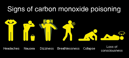

The clinical manifestations of CO poisoning can be subtle. Patients may be mistaken as having influenza (similar symptoms and both peak in winter months), presenting with headache, myalgias, confusion, tachypnea, tachycardia, presyncope and altered LOC. Classically, patients are described as having cherry red lips, though this is an insensitive sign. One retrospective study found the initial diagnosis to be listed as stroke, cardiac ischemia, encephalitis or seizure in multiple cases, highlighting the diagnostic challenge. Other acute symptoms include cardiac chest pain, and this has been described in 30% of cases. This is associated with increased mortality in CO poisoning. Chronic symptoms include the neuropsychiatric syndrome, which presents with personality changes, focal deficits, or cognitive impairment. This occurs with a month following poisoning and can persist for years. The mechanism is not clear, but may be related to myelin destruction from reactive oxygen species.

Because of the non-specific nature of CO poisoning a high index of suspicion is needed to make the diagnosis. The oxygen saturation on pulse oximetry is normal, given the maintained binding of hemoglobin. The diagnosis is made using co-oximetry to measure the amount of carboxyhemoglobin (venous or arterial blood). Amounts of carboxyhemoglobin can be found in several situations:

1-3% carboxyhemoglobin - Normal individuals
under 10% - smokers
Consider the patients history and presentation to determine if the levels are reasonable or as a result of poisoning.

The treatment of CO poisoning is emergency management.

1. ABC's - Many patients will have CO poisining from smoke inhalation. Security of the airway is crucial. Tachypnea, hypoventilation, stridor, facial blistering, burns and edema should prompt intubation and laryngoscopy. Patients can develop airway edema within 24 hours after the inhalation injury following thermal injury, this requires admission and monitoring for respiratory deterioration.

2. Oxygen therapy - all patients should receive high flow 100% oxygen. This results in rapid
improvement in CO levels compared to room air. The half life of CO is 300 minutes, and decreases to 90 minutes when supplimental oxygen is provided.

3. Consider hyperbaric oxygen therapy (HBOT) - Although this therapy is ubiquitous within the literature, the benefit of hyperbaric oxygen is somewhat controversial. There is no doubt that hyperbaric oxygen decreases the half life of oxygen even further than high FiO2 therapy, decreasing the half life to roughly 30 minutes. Whether this extends to improved long-term outcomes is debated. A cochrane review from 2011 found that further RCT's are required to determine whether HBOT should be offered, based on the poor quality of previous studies (see link below for details). Indications are variable, some directives suggests asymptomatic patients with a level of  over 40% CO should have HBOT, while others say 25% is a reasonable cutoff. Other reasons to initiate HBOT are:

Pregnancy and CO level over 20%
Decreased LOC or new neurologic deficits
Myocardial ischemia
Acidosis (less than pH 7.1)

4. Isocapneic hyperventilation - this is an experimental treatment that doesn't seem to be widely used in the clinical setting. One study (actually affiliated with anaesthesia at the University of Toronto), showed that in dogs, hyperventilating while providing inhaled CO2 to maintain pH balance resulted in improved removal of CO.

Cochrane review on HBOT


Wednesday, October 9, 2013

Rhabdomyolysis

Defined as dissolved skeletal muscle content, rhabdomyolysis is exactly that. It can present in many ways, from completely asymtpomatic to life-threatening end-organ damage.  In the early 1900's the syndrome of rhabdomyolysis was recognized as a cause of acute kidney injury following crush injuries. A case series corroborated this following the bombing of London in 1940, where patients injured from falling buildings were found to develop similar symptoms. Non-traumatic causes and the overall pathophysiology of rhabdomyolysis were not well recognized until decades later.


The final common pathway in causing rhabdomyolysis is increased intracellular calcium. Whether there is direct trauma to the sarcomere from trauma, or ischemia causing depleted ATP, all patients with rhabdomyolysis develop increased intracellular calcium. These leads to a worsening of intracellular energy stores, as Na/Ca exchangers actively use ATP to maintain normal electrolytes gradients. Increase reactive oxygen species leads to direct toxicity to cell membranes, and muscle death an intracellular content release occurs.

The causes of rhabdomyolysis are many, but can be broken down into several categories. I like the way its categorized on UptoDate, as it really allows you to structure your differential diagnosis. :

1. Traumatic - crush, trauma, immobolization from fall/coma (as in our patient today), burn, compartment syndrome

2. Non-traumatic exertional - significant exercise (marathon runner), myopathies that are exposed through exercise - (McCardle syndrome, carnitine palmitoyltransferasedeficiency)
Some people might include seizure in this category as it is a direct result of muscle contraction and exertion

3. Non-traumatic non-exertional - inflammatory myopathies, endocrine causes (hypothyroidism, DKA, pheo), infectious causes (viral, bacterial and parasitic causes - malaria), drugs (statin, alcohol, colchicine, herbal products, opioids, illicit drugs - cocaine) and electrolytes abnormalities (hypokalemia, hypophosphatemia)

The diagnosis can be suspected clinically, but we rely on laboratory findings to confirm rhabdomyolysis. The triad of weakness, dark urine and myalgias is typically present. On examination, muscles can appear swollen and tender on palpation. Additional urinalysis testing can be helpful. Myoglobinuria can be identified on dipstick at a level of 150mg/L, but can not be differentiated from hemoglobin. Microscopic examination will show minimal/no red blood cells if pure myoglobinuria. The pH of urine in this setting tends to be acidic. Proteinuria can be seen in up to 50% of cases. If there is more than 300mg/L of myoglobin in the urine, there will start to be a colour change to reddish/brown.

The main concern in managing rhabdomyolysis is the development of acute kidney injury. Its estimated that 10% of all AKI are related to rhabdomyolysis. However, the CK level doesn't correspond directly with increasing chance of AKI. That being said, renal injury usually develops at levels above 15,000. Mechanisms of renal injury include direct tubular toxicity, renal vscocrinstriction and tubule blockage due to myoglobinuria, not CK. This is interesting considering we measure CK levels, and not myoglobin levels in the serum (which are actually quite insensitive for the diagnosis if rhabdomyolysis). The direct tubular toxicity (mainly proximal tubule) is thought to be related to myoglobin biproducts, including free iron and production of reactive oxygen species, which can lead to direct tubular cell injury. Renal vasoconstriction is likely multi-factorial, given these patients are oven volume deplete, ill and reduced vasodilating agents (ie. NO). Tubular blockage occurs when myoglobin accumulates in distal tubules (most common). This process is enhanced by acidic urine, and volume depletion, which can cause increased Tamm-Horsfall protein casts with myoglobin can precipitate in.

Treatment is mainly focused on volume expansion to prevent AKI and treating the underlying cause. Certain IV fluids are promoted in the treatment for several reasons:

1. Normal saline - good volume expander if hypotensive/significantly dehydrated. Issues with this include the significant chloride load, which put patient at risk for metabolic acidosis. This may worsen some the physiologic mechanisms that can lead to myoglobin induced kidney injury (vasoconstriction, precipitation and tubular injury).
2. RL - reasonable choice given lack of chloride.
3. Sodium bicarbonate - will allow alkalinization of the urine, which will theoretically prevent the above issues with myoglobin. However, no RCTs have shown this strategy to improve outcomes. It seems reasonable to include this in therapy, especially if patients is hyperkalemic secondary to muscle injury.
4. Mannitol - suggested as a volume expander as it can lead to an osmotic diuresis and improved GFR, theoretically clearing tubules of myoglobin and cast debris.

Here is an article identifying some predictors for AKI in rhabdomyolysis.

Predictors of AKI and Rhabdo


Saturday, October 5, 2013

Cryoglobulinemia

Cryoglobulins are antibodies that precipitate at temperatures below 37 degrees Celsius. Its important to recognize that cryoglobulinemia is by definition the presence of precipitating proteins at low temperatures, however, clinicians tend to use this label to mean the clinical syndrome it is associated with. Cryoglbulins are classified into several categories, with each having its own clinical features and associations. :

Type 1 cryoglobulinemia - is monoclonal antibodies of one type, ie IgG or IgM. Associated with hematologic malignancies (Waldenstroms macroglobulinemia or myeloma). The primary clinical concern here is hyperviscosity syndrome where patients presents with infarcted digits, TIA/stroke like symptoms or thrombosis.

Type 2 cryoglobulinemia aka essential mixed cryoglobulinemia - results from monoclonal and polyclonal antibodies, including rheumatoid factor. The large majority of these patients have hepatitis C (30-100%), although connective tissue disease (Sjogren's syndrome) and lymphomas can also lead to this condition. Other less common infectious causes include HIV, streptococcal infections and brucellosis.

Type 3 cryoglobulinemia - is another form of mixed cryoglobulinemia, where all antibodies are polyclonal.

The classic triad was described by Meltzer in 1966, as purpura, arthritis and myalgia. These symptoms suggest cryoglobulinemic vasculitis. Palpable purpura is the most common manifestation, present in up to approximately 80% of patients. Purpura refers to bleeding under the skin that is larger than 3mm, when less than 3mm its termed petechiae. Other dermatologic findings include ulceration, levido reticularis and digital necrosis. Renal involvement is present in 30% of patients, where proteinuria, hematuria and active sediment production can occur. Nephrotic and nephritic syndrome occur in 21% and 14% of cases respectively. Neuropathy is also common, with up to 60% of patients describing symptoms burning and parasthesias. Mononeuritis multiplex (neuropathy involving a nerve with a name) is also seen, where patients can develop wrist or foot drop.

Testing for cryoglobulinemia is problematic, where samples need to be obtained in warmed syringes and kept at 37 degrees. Once serum is isolated it can be stored in a refrigerator at 4 degrees. It takes days for mixed immunoglobulin to precipitate, but type 1 proteins can be identified within hours. The quantity of cryoprecipitate can also be determined, using a test called the cryocrit. This is important because disease severity is proportional to amount of protein. Additional tests include complements, where low C4 is common, and elevated rheumatoid factor. Looking for an underlying disease association is also important if not already recognized.

Treatment is targeted towards the underlying cause and removing the monoclonal immunoglobulin. Considering HCV is so common in mixed cryoglobulinemia, ribavirin and interferon therapy should be considered with consultation by hepatology. Plasma exchange is helpful in life threatening disease with hyperviscosity. There is some growing evidence for the use of rituximab (monoclonal antibody targeting B-cells). A RCT from 2010 showed expedited rates of remission, improved renal involvement and higher rates of protein clearance compared to antiviral therapy in those with HCV. See a NEJM review on treatment in HCV associated cryoglobulinemia for more details (courtesy of the admitting physician).

HCV cryo treatment


Thursday, October 3, 2013

Heart block

Abnormal conduction between the atria and ventricles, termed AV conduction blocks, are common conditions in internal medicine. They range from mild asymptomatic findings to emergent, life threatening conditions. In 1964, Lev published the anatomic basis for atrioventricular blocks in the American Journal of Medicine, and still today, the disease that carries his name is responsible for nearly 50% of cases. The common nomenclature of AV block is documented as:

1st degree - slowed conduction between atria and ventricles with maintained synchrony
2nd degree - impaire conduction with intermittent loss of synchrony (separated into type I and II)
3rd degree - complete loss of synchrony between atria and ventricles. 

For the most part, conduction block are a disease of ageing or ischemia. Lev's disease, is used to describe the gradual fibrotic changes that occur with ageing to the cardiac conduction system, which lead to impaired functioning. Myocardial ischemia is the next most common cause, and can be acute or chronic. One study (Circulation, 1978) examined the natural history of heart block in patients following acute MI, and found that AV block is common. Twenty-two percent of patients developed second degree heart block type II. The most common type of blocks were LBBB and RBBB with anterior fascicular block. Patients who developed high grade AV block post MI had a significantly increased mortality (47% vs 23%), which was often directly related to the arrhythmia development and hemodynamic compromise. Other less common causes for AV block include:

Infection - endocarditis (aortic root abscess leading to AV pathway dysfunction), diptheria, scarlet fever, tuberculosis (with pericardial invovlement) mumps, lyme, toxoplasmosis
Inflammatory conditions - rheumatoid arthritis, lupus
Miscellaneous - sarcoidosis and amyloidosis (from infiltration)
Cardiomyopathy - hypertrophic cardiomyopathy, myocarditis'
Genetic - inherited forms of AV block exist, but are uncommon
Medications - remember to take a detailed history to identify agents that will impact the conduction system. 

Today, our patient was taking donepezil (an acetylcholinesterase inhibitor), which may have contributed to the development of bradycardia and heart block. There are multiple case reports of high grade AV block and even torsades des pointes with this class of medications. Donepezil is thought to be more associated with SA node dysfunction, causing sinus bradycardia more than high grade AV block, but both have been documented.  

After treating possible underlying conditions and removing offending medications, the next step is to consider pacing and pacemaker insertion.Determining treatment for the large majority of patients, which have ischemic or age related conduction disease, is based on symptoms and risk of worsening conduction delay.  
Generally accepted reasons for pacemaker insertion in AV block include:

1. Complete heart block, 
2. Symptomatic second degree heart block
3. Second degree block type II with consecutive dropped atrial contractions
4. Second degree block with previous wide complex on ECG
5. Exercise induced high grade blocks

Patients with bifascicular and trifascicular block may also require a pacemaker in the setting of syncope, given it may be due to the development of transient third degree heart block. More detailed instructions can be found in the AHA guidelines for pacemaker insertion linked below.


Wednesday, October 2, 2013

Enterococcus and endocarditis

Infective endocarditis (IE) is a huge topic. The IDSA guidelines are very comprehensive and cover the treatment of IE based on microbiology and valve involved (mechanical vs bioprosthetic). These are linked below.

Enterococcus fecaelis and fecium were given their name to emphasize their presence in the human gastrointestinal tract. There are multiple species of enterococcus, but these two tend to be the most clinically relevant bugs. In the 1930's, the Lancefield classification placed enterococcus in group D, along with strep gallolyticum. Enterococcus was identified as a cause for IE as ealy as 1906, and over the following decades found to cause urinary tract infections, biliary infections, peurperal sepsis, and wound infections during WWI. 

Infective endocarditis is caused by enterococcus in 5-15% of cases. The virulence of this organism is much lower compared to other organisms (ie. S. aureus). Two studies from the 1980's found that bacteremia from enterococcus had a relatively low likelihood of developing endocarditis, where only 2.5% of those with positive blood cultures met criteria. E. fecaelis is more common than E. fecium, which often requires vancomycin therapy given penicillin resistance. Even less frequent than this is vancomycin resistant enterococcus as a cause for endocarditis.  Enterococcal endocarditis is more common in men and older patients, where the average age in one study was 65.  One study from the archives on internal medicine showed that 50% of cases in men followed urinary tract instrumentation, an important risk factor that was present in our case discussion. Other associations include a history of malignancy and nosocomial acquisition. Below is a relatively recent retroprospective database from the American Jounral of Medicine.

Anecdotally, enterococcal endocarditis is less commonly associated with peripheral stigmata, which were absent in our patient. The disease course tends to be subacute with aortic valve involvement being most common.  

Unfortunately, enterococci are less susceptible to beta-lactam antibiotics and synergy with aminoglycosides are often employed. The purpose of dual therapy is to have cell wall breakdown from the penicillin antibiotic allow for improved penetration of the aminoglycosides, which act intra-cellularly on ribosomal activity. Different strains can have varying susceptibility to these antibiotics, which may influence dosing and antibiotics choice. Gentamicin/streptomycin have toxic effects, including ototoxicity and nephrotoxocity and the minimum six weeks of antibiotics required for this diagnosis can lead to significant morbidity. Prosthetic valve endocarditis may fail to be effectively treated with antimicrobials alone, and often requires surgical opinion. I would also advocate for the involvement of the ID service to help with antibiotic choice, dose, interval and duration of therapy.

Tuesday, October 1, 2013

Chronic myeloid disorders

Chronic myeloid disorders can be a confusing group of diseases to understand. There is overlap between conditions, making diagnosis challenging at times and many conditions have multiple names. Today we discussed a case of myelodysplastic syndrome, which is very likely a case of myelofibrosis. Here, we describe a general approach to these conditions and some of the clinical features.

Chronic myeloid disorders is an umbrella term encompassing multiple conditions: chronic myeloid leukemia (CML), myelodysplastic syndrome (MDS), chronic myeloproliferative disease and atypical chronic myeloid disorders. Chronic myeloproliferative disorders can be broken into polycythemia vera (PRV), essential thrombocytosis (ET) and idiopathic myelofibrosis (IMF). I think that confusion arises as a result of the multiple subgroups within each category of disease. You need to read about each entity separately to gain a better understanding of its differences.

MDS is as a result of ineffective and disordered hematopoeisis in one or more cell lines. Despite low peripheral counts, bone marrow evaluation reveals normal or hypercellularity. This is generally a disease of the elderly, where the majority of patients are over 65. It can be seen earlier, increasingly if caused by an external chemical exposure (ex. benzene). Clinical features are those associated with low blood counts; infection, bruising, fatigue etc. Some additional hints towards a diagnosis include an increased number of band neutrophils on the smear, the so called "pseudo-Pelger-Huet" anomaly, a result of premature cells being pushed into the blood.

The classification of myelodysplastic syndromes (MDS) changed in 2001. The World Health Organization changed the previous French-American-British (FAB) definition to improve the diagnosis of AML. MDS are a group of myeloid neoplasms, that impact on the proliferation of hematopoietic cells. Patients can present in a myriad of ways, and are generally classified into multiple categories:

1. Refractory cytopenia with unilineage dyslpasia- low RBCs or low platelets or low neutrophils
2. Refractory cytopenia with multilineage dysplasia- multiple low cell lines
3. Anemia with ringed sideroblasts- sideroblasts are abnormal mitochondria that are visibly different as a result of increased iron content
4. Refractory anemia with excess blasts - two stages based on percentage blasts (5-10%, 10-20%)
5. MDS from 5q deletion- a specific genetic variant
6. MDS otherwise unclassified

Prognosis varies depending on the subtype of MDS. Twenty percent of patients with MDS will die from AML, however those with 5q deletion are less likely to convert, and have a more indolent course.

Myelofibrosis (IMF), aka agnogenic myeloid metaplasia, is a different disease. This is a clonal stem cell disorder with progressive bone marrow fibrosis. An abnormal precursor is thought to produce factors that result in fibroblast proliferation and collagen deposition. Premature fibrosis pushes precursor cells into the periphery resulting in deposition of these cells in distant tissues. This leads to hematopoeisis outside the marrow. Cells can deposit in many different organs, including the lungs, pericardium, spleen, bowel, liver etc. IMF is less common than MDS, present in only 0.5 people per 100,000. The clinical presentation includes fatigue, splenomegaly, constitutional symptoms, and complications of bone marrow failure. Counts are often increased initially because of compensatory extramedullary hematopoeisis. Blood smear will often show premature WBCs, teardrop cells, giant platelets, termed a leukoerythroblastic pattern. Up to 60% of these patients will have a testable mutation in a gene called JAK-2. Occasionally,  can be difficult to distinguish IMF, from burnt out ET or PRV.

The prognosis in IMF is poor, with 10% of patients transforming to AML, having an average survival of 2-5 years. Unfortunately there are few successful treatments for this condition. In young individuals, allogeneic stem cell transplant can be considered, but in the elderly there is little that can be done. Blood counts can be controlled with various chemotherapy, and low counts managed with transfusion.

Two reviews for each of these conditions are listed below.

MDS review - NEJM
Myelofibrosis and NEJM