Thursday, December 17, 2009

Phenytoin (Dilantin) toxicity


Yesterday we discussed dilantin toxicity. Dilantin is an anticonvulsant that can cause a number of drug reactions of different types:

Pharmacology:
Phenytoin binds to and inhibits sodium channels in neurons and in cardiac tissue

It is cleared by the liver via the CYP450 system.

An important clinical point about phenytoin pharmacokinetics is that it exhibits "zero-order" kinetics. This means that only a fixed amount (not proportion) of drug is metabolized after a certain point (which is unknown for a given patient). If this threshold is crossed, a very small increase in dose can cause a big increase in level and toxicity. Increase doses slowly and by small increments (e.g. 25-50mg/d at a time, checking levels).

There are many drugs that can increase and decrease phenytoin levels via CYP450 interections. Click here for a complete list.

Phenytoin toxicity ("poisoning")
The earliest sign is nystagmus (usually horizontal) and unsteady gait. More severe toxicity causes slurred speech, lethargy, confusion, and eventually coma.
It can rarely cause cardiac arrhythmias (mainly bradycardia, AV blocks, sinus arrest)
There is no specific antidote for phenytoin; treatment is supportive.

Chronic effects/toxicity
Neurological involvement as above, gingival hyperplasia

Idiosyncratic reactions
These are non-dose related effects.

Drug hypersenitivity syndrome
Characterized by fever, rash (with or without mucosal involvement), and internal organ involvement. Sometimes also called "DRESS" or "drug reaction with eosinophilia and systemic symptoms". Dilantin is a rare, but very well described culprit, along with sulfonamides, allopurinol, dapsone, and many others. Timeframe is 2-8 wks after initiation.

Stevens-Johnson's, Toxic Epidermal Necrolysis
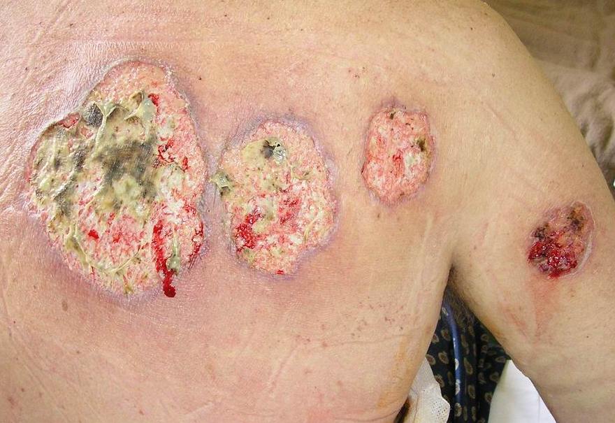
Desquamating skin and mucosal involvement; organ failure. Distinguished by surface area involved. Less than 30% BSA = SJS. More than 50% = TEN; overlap = between. Dilantin is a well described culprit. Tx: supportive (inc. burn unit), possible role for steroids, IVIG.

Drug-induced lupus
Clinically, mainly arthritis, serositis, wt loss. 95% have anti-histone AB, negative anti dsDNA, normal complements.

Others
Isolated hepatitis
Leukopenia, thromboctyopenia, agranulocytosis
Lymphadenopathy
IV preparation can cause hypotension during infusion (treatment is fluid)

Links
Click
here for a good overview of idiosyncratic drug reactions
Click here for a summary of phenytoin kinetics

We'd
like to know who reads Tangents, and what you think of this blog. Please click here to take a short survey. Thanks!

Tuesday, December 15, 2009

Hepatic encephalopathy


Today we discussed hepatic encephalopathy and its triggers. Some points that came up:

In the context of stable chronic liver disease, the triggers of hepatic encephalopathy are:
1) non-adherence to diuretics/lactulose/nadolol, etc.
2) psychoactive meds (narcotics, benzos)
3) volume overload (usu insufficient diuretics/NaCl excess)
4) new hepatic insult: hepatitis, HCC, PV thrombosis, Budd Chiari
5) infection: esp SBP
6) GI bleed/protein load
7) hypokalemia/alkalosis

Approach is to control encephalopathy and rule out/in precipitating causes:
1) All cirrhotics with ascites and worsening encephalopathy must be tapped. Also rule out other causes of infection
2) Lactulose 30cc BID, qid PRN for 2-3 loose BMs.
3) Diurese if needed, but consider holding if intravascularly depleted
4) D/C all meds that could contribute
5) Continue/start nadolol
6) Decide on ABx from clinical picture and preliminary tap cell count
7) Consider U/S with dopplers to r/o PV thrombosis, HCC, etc
8) If suspicion of GIB, possible OGD
9) Low salt diet. Fluid restrict.

Click here for the 2004 guidelines for cirrhosis managment from Hepatology

We'd like to know who reads Tangents, and what you think of this blog.
Please click here to take a short survey. Thanks!

Monday, December 14, 2009

Subclinical hyperthyroidism


Today we discussed subclinical hyperthyroidism


This is defined as
1) Low (usually undetectable) TSH
2) Normal T3 and T4
3) no symptoms of hyperthyroidism

The causes of subclinical hyperthyroidism mirror the causes of overt hyperthyroidism:
1) Exogenous T4
2) Toxic adenoma or multinodular goiter
3) Subacute thyroiditis
4) Graves' disease
5) Amiodarone
Others...

Differential of a low TSH with normal T3/T4 besides subclinical hyperthyroidism:
1) "Sick euthyroid"- stress causing decreased peripheral conversion to T3 (i.e. low), normal T4, low TSH.
2) Glucocorticoids, dopamine
3) Autonomous thyroid nodule producing enough T3/T4 to suppress TSH but not cause high T3/T4 levels

Workup consists of same as overt hyperthyroidism: radioactive iodine uptake and scan, possibly thyroid-stimulating antibodies.

Concerns in subclinical hyperthyroidism
1) Atrial fibrillation
2) Osteoporosis
3) Risk of progression to overt hyperthyroidism
4) Neuropsychiatric effects, diastolic dysfunction (softer evidence)

Natural history: Depends on underlying cause; ~2-5%/yr progress to overt hyperthyroidism

Whom to treat?
Controversial! Very little evidence.

This structured review from JAMA suggests considering treatment for patients who are older than 60 years and for those with or at increased risk for heart disease, osteopenia, or osteoporosis, or for those with subclinical Graves disease or nodular thyroid disease

Therapy consists of ablative therapy (i.e. radioactive iodine) with replacement or medications (PTU or methimazole) with goal to normalize TSH.

We'd like to know who reads Tangents, and what you think of this blog. Please click here to take a short survey. Thanks!

Saturday, December 12, 2009

Inhalational injury


Today we discussed inhalational injury.

This is suggested by:
-soot in the upper airway, including mouth and nares
-singed eyebrows, nares
-persistent cough, stridor, wheezing
-hoarseness

Some principles of managment:
1) Consider early intubation; peak of airway edema is 48-72h after injury, and may make intubation extremely difficult. ARDS is also common following thermal injury
2) Look for carbon monoxide poisoning (more below)
3) Bronchoscopy should be performed initially then frequently to assess the extent of the injury and to clear sloughed airway epithelium
4) Look for cyanide poisoning (from plastics and fabrics burning). This presents as a triad of altered mental status, cardiovascular collapse, and lactic acidosis
5) Ophthalmology should see patients with inhalational injuries to exclude ocular injury, since they often go together

A few points about carbon monoxide poisoning:
Presentation
-Notoriously difficult to diagnose
-Common presenting symptoms are altered mental status, headache
-Physical exam may show a "cherry red hue" but this is unreliable

Labs
-Oxygen saturation is normal! (oximeter reads carboxygemoglobin as oxygen-bound)
-ABG tends to be normal (PO2 is dissolved oxygen, which is not changed by CO)
-Need to do co-oximetry on ABG sample to detect carboxyhemoglobin (reported as a percentage)

Management
-100% oxygen by non-rebreather mask for everyone (decreases the halflife of carboxyHgb from 300 to 100 min)
-Hyperbaric oxygen therapy decreases the halflife to 30 min. Generally recommended for any of 1) Carboxyhemoglobin level over 25% (although some sources suggest 4o%) 2) Signs of ongoing organ ischemia (lactic acidosis, myocardial ischemia, etc) 3) Loss of consciousness, 4) Pregnancy with a level over 20% or fetal distress

Links
Click here for a Cochrane review of hyperbaric oxygen in CO poisoning by Toronto investigators (including this hospital!)
Click here for a website from the University of Utah outlining burn management

We'd like to know who reads Tangents, and what you think of this blog.
Please click here to take a short survey. Thanks!


Wednesday, December 9, 2009

Psychosis: medical causes


Today we discussed the approach to the psychotic patient from a medical standpoint (i.e. causes other than primary psychiatric disease)

See this post for a general approach to dementia.

In terms of psychosis (i.e. hallucinations or delusions) from a medical cause, things to think about include:

Delirium
Hallmark is attention problems. Also cognitive problems, simple hallucinations (often visual), fluctuating course, underlying medical condition
General causes:
Infection, intoxication (esp. anticholinergic; 'mad as a hatter'), medications (e.g. benzos, opiates), substance withdrawal (esp. alcohol), metabolic derangements (Na, Ca, hepatic encephalopathy, uremia), trauma, CNS pathology, hypoxia, deficiencies, endocrine (e.g. hyper or hypothyroidism, hypoglycemia), vascular (e.g. vasculitis, hypertensive emergencies)

With more elaborate hallucinations/delusions, other medical possibilities that are well known to present with psychosis include
-Seizures (especially temporal lobe)
-Encephalitis (especially HSV encephalitis)
-Brain tumor / bleed involving the temporal lobe
-Lewy Body dementia (classically non-bizzare delusions, worsens with neuroleptics)
-Neurosyphillis
-Wilson's disease
-Hyperthyroidism
-Neuropsychiatric lupus
-Carbon monoxide poisoning
-Charles Bonnet syndrome
(complex visual hallucinations with insight in pts with visual impairment that reverse with visual correction)

-"megaloblastic madness" from B12 or folate deficiency
-Porphyria

Link:
Click here for a summary of medical causes of psychosis to consider.

We'd like to know who reads Tangents, and what you think of this blog.
Please click here to take a short survey. Thanks!

Monday, December 7, 2009

Hypoglycemia practical management


Today we discussed hypoglycemia in the context of medications.

For a discussion of the differential diagnosis of hypoglycemia, see

this post

Some considerations for dealing with hypoglycemia on the wards:

2 issues: 1. Increase blood glucose. 2. Determine reason

Gucose is below 4: If patient is awake, not NPO,

1. Need 15g carbohydrate (in the form of glucose tabs, 3 packs table sugar in water, 3/4 cup of juice or regular soft drink, 6 life savers)If BG is below 2, need at least 20g glucose.

2. Check glucose q 15 min until above 5, and repeat 15g CHO as needed

3. If over 1h until next meal, snack (e.g. 1/2 sandwich)

4. Look for cause, address.

5. Do not stop insulin in a type 1 diabetic


Pt NPO:
1. 25g CHO in form of D50 (50mL amp) push or D10 500cc. May also give glucagon 1mg sc/im if delay.

2. Check glucose q15 min until it is over 5 and give more D50 or D10 as needed

3. Pt may need ongoing glucose infusion after (e.g. D5 or 2/3+1/3 or D10)

4. Look for cause

5. Do not stop insulin in a type 1 diabetic

Reasons: Insulin or sulfonylurea excess or decreased PO intake.

NB- in sulfonylurea-induced hypoglycemia, consider octreotide as reviewed

here


Friday, December 4, 2009

Pulmonary hypertension


Today we discussed pulmonary hypertension (or, we at least had a "tangent"... on it.)
This is an important clinical entitiy to have an approach to both in terms of physical findings and the workup required to determine the etiology. Some considerations:

Defined as mean PA pressure over 25mmHg at rest with mean LA or wedge pressure below 15 (i.e. no LV dysfunction)

May be primary (idiopathic) or secondary.

May occur from disease anywhere in the following circuit:
PA, lungs, PV, LA, MV, LV

1) Pulmonary artery- luminal (PE), mural (PPH), extramural (tumor)
Clinically and patologically indistinguishible from primary pulm HTN are cases associated with the following: anorexigens (diet pills), cocaine, HIV, rheumatologic diseases (esp CREST/scleroderma), portal HTN

2) Lungs- chronic obstructive and restrictive diseases can cause PHTN through hypoxic vasoconstriction. Interstitial lung disease can also cause destructive changes to pulmonary vasculature, leading to PHTN and cor pulmonale.

3) PV- veno-occlusive disease

4) Cardiac- L CHF- usually causes relatively minor PHTN. Mitral stenosis can cause severe PHTN. Shunts (ASD, VSD) cause higher R-sided pressures. Increased PA flows cause changes in PA structure and subsequent reversal of flow (Eisenmenger's)

Clinical features:

Hx: Sx of heart or lung disease, HIV RFs, Sx suggestive of CTD, esp scleroderma, anorexigens, PE symptoms, Fhx (in 5% of pphtn), snoring/OSA, rheumatic heart disease

P: Loud P2, RV lift (L parasternal), R-sided S4, TR murmur (holosystolic, louder on inspiration), edema, high JVP. A murmur of pulmonic insufficiency called the "Graham Steell murmur" is described (early diastolic, decrescendo, increased on inspiration). Look for findings suggestive of secondary causes including scleroderma, HIV, primary lung disease, etc.

Investigations:
ECG- RAE, RVH with strain (i.e. tall R and small S with R/S over 1 in V1)
CXR- enlarged R hilum, dilated PA (over 14mm in females, over16mm in males)
Echo- RVSP (approximaes PA systolic). Should be less than 40.
PFT/ABG
High resolution CT +/- VQ scan
Possible R heart cath to determine pressures directly


Link:
Click here for an excellent JAMA "grand rounds" discussion on pulmonary HTN

Wednesday, December 2, 2009

Essential thrombocytosis


Today we discussed essential thrombocytosis (AKA essential thrombocythemia). Some points about this condition:

Thrombocytosis
Majority of cases are reactive. Common causes: Post-op, infection, post-splenectomy, Fe deficiency, malignancy (non-hematologic)

Non-reactive causes include ET, polycythemia vera, myelofibrosis, CML, MDS

ET:
Concerns are
1)Thrombosis risk
2)Bleeding risk (platelets are numerous, but often dysfunctional, and patients can have acquired von willebrand's disease)
3)Potential for transformation to AML


Epidemiology:

median age at dx is 60. F:M is 2:1.
Clinical: ~1/3 asymptomatic. ~1/3 have "vasomotor" sx: Syncope/presyncope, H/A, paresthesias, visual dist, non-exertional c/p. ~1/3 have sx consistent with thrombotic/bleeding events- major or minor

Diagnosis:

PLT consistently above 600, no explanation for reactive cause. May see megakaryocyte hyperplasia on BMBx if done, -ve BCR-ABL, iron replete, no evidence of MDS. JAK2 is a mutation found in over 95% of patients with polycythemia rubra vera, and 50-60% of patients with ET. It can be detected in peripheral blood

Bleeding risk
This paper by Tefferi et al from the Mayo Clinic found a 4% 10-year risk of major hemorrhage. Bleeding risk was directly proportional to PLT level, with over 1500 (i.e. 1.5 million) highest.

Thrombosis Risk
Arterial and venous thrombosis may occur. Typical events are similar to those in PRV: stroke, TIA, ACS, retinal art occ, digital ischemia, pregnancy loss. Less commonly, DVT/PE, PV thromb/Budd-Chiari.
Incidence of major events (MI, stroke, PE) is ~4% at 10 years. There is conflicting data on whether PLT level correlates with thrombosis risk


Antiplatelet therapy
The ECLAP study (RCT) looked at low dose ASA in patients with no other indication for ASA and PRV (which carries similar thrombosis risk)
RR of composite outcome of MI, stroke, PE, CV death was 0.4 on ASA 100mg. No significant mortality benefit. No significant difference in bleeding. It also has demonstrated effect on vasomotor sx of ET.


Cytoreductive therapy in high risk pts
In those meeting high risk criteria (i.e. over 60
or history of thrombosis). this RCT by Cortelazzo et al, evaluated the use of hydroxyurea to bring PLT count to below 600 vs. placebo in patients on antiplatelet therapy. 2 year risk of significant thrombosis was 3.6% on HU, 24% on placebo (NNT 5) Therefore, hydroxyurea to reduce the platelet count to below 600 should be considered in high risk patients. Specific platelet reducing agent (Anagrelide) was evaluated in RCT vs. HU, and showed much higher thrombosis risk


The risk of conversion to AML is reported at 2-5% at 15 years from dx. This is the lowest risk of any of the myeloproliferative disorders

Monday, November 30, 2009

Hypertensive emergency


Today we discussed hypertensive emergencies. Some important points:

Some of the confusing terminology in this area:
"Urgency": SBP over ~180 or DBP over ~110 without end-organ damage- needs correction over days with oral agents
"Emergency": Above, but with acute end-organ damage, needing urgent lowering, usually with IV medications in a monitored setting.
"Malignant": Old term referring to HTN with encephalopathy or renal involvement
"crisis", "accelerated"- largely abandoned, and replaced with "emergency"


End organ complications and specific treatments:
1) Aorta- dissection (B-blockade, nitroprusside after B-bl. No pure vasodilators)
2) Brain- encephalopathy- sz, coma (avoid centrally acting agents); cerebral hemorrhage/infarction, raised ICP
3) Heart- MI, CHF (acute diastolic dysfunction leading to pulmonary edema)- careful with B-bl. May use nitro infusion
4) Kidney- renal failure- careful diuresis, calcium antagonists useful
5) Placenta (pre-eclampsia)- hydralazine, labetalol, delivery
6) Hemolysis (can look just like TTP with MAHA, fragments)


Emergency pts should go to ICU, and have art line. Generally, most extra-cerebral damage benefit from rapid lowering.

Aim for MAP decrease by no more than 20-25% within 2h or to DBP 100-110 over minutes to hours. Exceptions include aortic dissection, where more rapid decrease may be needed, and stoke, where less rapid lowering may be needed.

Special circumstances:
Cocaine- phentolamine or labetalol (avoid unopposed alpha-agonism)
Pheochromocytoma- phentolamine/phenoxybenzamine (as above)


Drugs for acute lowering:
Labetalol 10-20mg bolus then infusion
Hydralazine- 10-20mg bolus (causes reflex tachycardia)
Nitroglycerine 5-100mcg/min
Na nitroprusside- arterial vasodilator
Phentolamine -alpha antagonist


Available in some centres:
Enalaprilat (IV ACE-I)
Fendolapam (arterial vasodilator)

Link:

Click here for a good review on this topic from Chest

Sunday, November 29, 2009

TTP


We discussed Thrombotic Thrombocytopenic Purpura (TTP), a very important condition to recognize and institute treatement because of its 90% untreated mortality.

Classic pentad:
1) Thrombocytopenia
2) Microangiopathic hemolytic anemia (MAHA; recognized by hemolysis with RBC fragments-shown above)
3) Neurological signs/symptoms
4) Renal failure
5) Fever

HOWEVER,
the full pentad is seen late in the course. Thrombocytopenia and MAHA without another cause is generally enough to start therapy

Pathologically, thrombin/platelet depositions in organs.

Pathogenesis: ADAMTS13 is a metalloproteinase which degrades von Willebrand's factor. Loss of this enzyme (acquired or genetic) causes large multimers of vWF to accumulate, activating platelets and causing microvascular clotting.

Causes of acquired TTP:
Infection: E.Coli 0157:H7, which causes colonic vascular injury and may induce TTP.
Drugs: ticlopidine, clopidogrel, cyclosporin
Cancer: TTP is assoc with Br, GI, pancreatic, prostate
Autoimmune disease: SLE (most common), RA, dermatomyositis, others.


Treatment:
Plasma exchange: Deficiency in ADAMTS13 is corrected, and antibody against it is removed. Done daily until LDH and PLT normalize. Large volumes of FFP may be transfused until transfer to a PLEX centre

Steroids- prednisone 1mg/kg/d PO or solumedrol 125mg IV BID as adjunct to PLEX.

PLT transfusion- not recommended unless life-threatening bleeding

Links:

Click here for the NEJM paper from Ottawa first showing the substantial benefit of PLEX

Click here for a NEJM review article on TTP

Thursday, November 26, 2009

Tricuspid Regurgitation


For sportscar connaisseurs, the iconic Ferrari 512TR

Today we discussed tricuspid regurgitation at physical exam rounds. Some points:

There are generally 2 classes of TR: 1) High pressure, usually secondary to L-heart disease (high RVSP), and 2) low pressure, usually from bacterial endocarditis.

In general, only the high pressure variety is detectable on physical exam because a significant pressure gradient between the RV and RA is required to generate the findings listed below.

The JVP:
-Elevated JVP: Seen in 90% of patients. Its absence strongly argues against high pressure TR
-CV waves: a systolic impulse of the neck veins is seen in about 50-80% of patients. You normally expect to see a descent in the JVP (the X' descent) during early systole (i.e. during and right after S1). If you do not see a descent, the patient probably has a CV wave.


Precordial palpation:
-If the RV is dilated, it may occupy the space where the LV normally lies (i.e. the apex). In this situation, you may see and palpate a systolic retraction of the apex with an outward movement of the L or R lower sternal borders (where the dilated RA lies), which is described as a "rocking" motion

Murmur:
-The murmur of TR is holosystolic. In 75% of patients, it becomes louder with inspiration (called Carvallo's sign). It is usually loudest at the L lower sternal border, but if the RV is dilated enough may be loudest at the apex. The LR+ for a typical murmur is 14.6. However, the lack of a typical murmur does not rule out TR (negative LR 0.8 for mild, 0.4 for severe).

Other:
-Pulsatile liver may be palpated (wide range of sensitivities reported). It is not 100% specific for TR (constrictive pericarditis and hepatic AVMs may also cause it), but by far the most common cause. Its presence argues that the TR is moderate to severe.
-Edema, ascites: 90% of patients have edema or ascites (or both)


Reference:
There is no JAMA RCE specifically for TR; most of above is taken from
Evidence-Based Physical Diagnosis (McKee)


Tuesday, November 24, 2009

Inflammatory bowel disease


Today we discussed inflammatory bowel disease. Some important points:


Extraintestinal manifestations of IBD:
Eyes- uveitis
Skin-
pyoderma gangrenosum, erythema nodosum

GI- PSC (esp UC)
Renal (stones- affects oxalate metabolism by unabsorbed bile salts binding Ca, allowing oxalate absorption)
Arthritis- Sero-ve, large joint asymmetric. Either peripheral, which parallels IBD or independent (axial)


Extent of UC (treatment):
pancolitis (oral meds), L sided (enemas), sigmoiditis/proctitis (suppositories)

Complications:
UC- bleeding, colon ca, toxic megacolon
Crohn's- aphthous ulcers, malabsorption, fistulae, abscess, strictures
Both: At higher risk for C. Diff, relatively hypercoagulable


Toxic megacolon: Non-obstructing dilation of the colon.
NB- TM can be from infectious causes as well.
Clinical: tachy, hypotension, fever, volume depletion, altered sensorium.
AXR: over 6cm at transverse, thumbprinting (big haustra), pneumatosis coli. Shown in above picture.

Flare treatment:
NPO (bowel rest)
NG
fluid resuscitation (esp attention to phos, K, Mg, albumin)
Possible antibiotics (esp. Crohn's) -cipro, flagyl, possibly vanco
IV steroids (e.g. Solumedrol 30mg IV bid)
DVT prophylaxis
Possible c-scope, although mucosa is very friable; may be deferred until inflammation settles.

Links:

Click here for a recent BMJ review of Crohn's disease

Click here for a Lancet review of new therapies for IBD

Monday, November 23, 2009

Pneumocystis jirovecii pneumonia


PCP (now PJP) is an opportunistic infection commonly seen in HIV positive patients with CD4 counts below 200. Ubiquitous in the environment, but it is very uncommon for immunocompetent patients to be infected.

Besides HIV, the other major risk factor is steroid use. In general, after 2 weeks of corticosteroid use at 20mg or more, PCP prophylaxis is indicated.

The classic clinical presentation is subacute onset dyspnea and dry cough. Other signs may include fever, tachycardia, and tachypnea. The chest exam is variable - there may be crackles, but in up to 50% of cases, the respiratory exam is normal.

The chest X-ray often reveals bilateral interstitial infiltrates, but virtually any abnormality may be seen.

Remember that PCP is a classic example of "CXR-negative pneumonia", where the immune reaction necessary to generate an infiltrate is absent. Pneumothorax may complicate PCP (the c is for cysts, which may rupture into the pleural space). Click here for a CXR showing this

Confirmation of diagnosis:
Silver staining of induced sputum (if possible) or BAL specimen (gold standard). PCR is under investigation.


Therapy:

1) TMP-SMX in high doses. In the event of sulfa allergy (interestingly more common in the HIV population), other agents can be used, such as pentamidine, dapsone, or atovaquone.
With high-dose TMP-SMX, look out for hyperkalemia, renal failure, hypoglycemia

2) Steroids for PO2 of 70 or lower or A-a gradient of 35 or higher.

Regimen is a 21-day oral taper of prednisone. The research showing the benefit of steroids in PCP comes from Toronto, and Toronto Western was part of the trial.


Other links:
Click here for the paper discussed by Marrie et al on "atypical pneumonia" and how clinical features do not reliably distinguish "typical" from "atypical" organisms.

SPECIAL NOTE:

Please click here for a Globe and Mail article about our very own Dr. Ho Ping Kong and his legendary morning report!


Thursday, November 19, 2009

Obstructive lung disease


At physical exam rounds, we discussed the diagnosis of obstructive lung disease.

There are many potential findings, outlined below, and 3 major papers on the evidence-based diagnosis of obstructive lung disease.


Possible findings:

General inspection:
Signs of respiratory distress (accessory muscle use, indrawing, paradoxical abdominal movement), pursed lip breathing, barrel chest, signs of malnutrition, look for clubbing (not expected in COPD), asterixis from CO2 retention, cyanosis, elevated JVP from cor pulmonale, many other possibilities...

Vitals:
Pulsus paradoxus

Palpation:
Subxiphoid cardiac impulse, palpable P2 from pulmonary HTN

Percussion:
Hyperresonance
Decreased diaphragmatic excursion
Decreased cardiac dullness


Auscultation:
Wheezes

Special manouevers:
Forced expiratory time (Patient takes a deep breath and exhales forcefully with open mouth, and examiner listens over lower trachea)
Laryngeal height- measure the maximum distance between the sternal notch and the thyroid cartilage. Less than 4cm is significant.

Evidence:

From JAMA Rational Clinical exam (1995):

Most sensitive tests (i.e. rule out if not present)- no single test sensitive enough
Most specific tests (i.e. rule in if present)
Wheezing (LR 36)
Barrel chest (LR 10)
Decreased cardiac dullness (LR 10)
Match test (patient unable to blow out match held 10cm in front with open mouth) (LR 7.1)
Hyperresonance
Forced exp time over 9 seconds
Other less useful tests to rule in, but positive LR's: Forced exp time 6-9s, subxiphoid impulse, pulsus paradoxus over 15, decreased breath sounds.


From JGIM- Straus et al, 2002
Took 161 pts with varying disease severity (known, suspected, or no COPD), did spirometry on all, looked at components of history and physical that predicted FEV1 5th percentile.

Key point here is combining findings is powerful in ruling in or ruling out.

Forced exp time over 9 seconds - LR 6.7
Wheezing - LR 4.0
Self-reported COPD LR 5.6


If all 3, LR 59 (rules in). If none, LR 0.3 (i.e. good for ruling out)

Other significant features: Over 40 pack-year smoker: LR 3.3


From JAMA- Straus et al (2000)- primary study, not Rational Clinical Exam

History of smoking over 40 pack-years: LR 8.3
Self-reported COPD: LR 7.3
Maximum laryngeal height less than 4cm LR: 2.8
Age over 45 LR 1.3

If all 4, LR is over 200
If none, LR- is 0.13

Links

For JAMA Rational Clinical Exam abstract click here

For Straus et al JAMA paper click here

For Straus et al JGIM paper click here


Tuesday, November 17, 2009

Glomerulonephritis


Today we discussed glomerulonephritis

GN is suggested by hematuria, proteinuria, HTN, edema. The hallmark is RBC casts on urinalysis

First division of GN is primary vs. secondary.

Secondary is suggested by associated symptoms and signs: fever, arthritis, rash
bloodwork for secondary causes includes ANA, complements, dsDNA, ANCA, HBV, HCV, HIV, SPEP, cryoglobulins, rheumatoid factor

Primary GN is divided into proliferative and non-proliferative.

Non-proliferative means no extra cells in glomerulus; presents as nephrotic end of spectrum.
These are generally less aggressive diseases:

1) Minimal change 2) Membranous 3) FSGS

Minimal change- mainly children, but seen in adults too. Rapid onset and offset of nephrotic syndrome. Tx is prednisone

Membranous- Most common primary GN in white males. Gradual onset of nephrotic syndrome. Associated with solid tumors in males over 60. Treatment is controversial.

FSGS- second most common primary GN ~40% respond to high dose prednisone

Proliferative GN- Means increased cells in glomerulus. Prominent RBC casts, hematuria, 'nephritic' presentation. Generally requires more aggressive immunosuppressive treatment

IgA nephropathy:
usually asymptomatic. Sometimes presents as post-URTI hematuria (within 1-5d). Rarely, rapidly progressive. 20% will progress; becomes relatively common reason for ESRD. Treatment is controversial; steroids, fish oil, ACE, ARB

Post-infectious (AKA post-streptococcal). Follows infection by 10-21d. Tx is supportive; usually self-limited. Possible in adults, but less common than children

Crescentic:
may be called "rapidly progressive GN" (although other types of GN can progress rapidly)
Divided by immunofluorescence findings:
1) pauci-immune (means ANCA +ve Wegener's, Churg-Strauss, others).
2) immune complex (vasculitis, endocarditis, SLE)
3) linear (Goodpasture's or anti-GBM)
Tx here is aggressive, intensive immunotherapy. PLEX for anti-GBM.


Post-infectious complications of streptococcal infection
Acute rheumatic fever
Glomerulonephritis
Reactive arthritis
Erythema nodosum

Links:

Click here for a review article on IgA nephropathy from the Toronto Nephrology Rounds series

Click here for a NEJM clinical pathological conference addressing a practical approach to renal failure

Sunday, November 15, 2009

Febrile neutropenia


Febrile neutropenia is a common condition that requires an organized approach and awareness of how this situation differs from fever in the immunocompetent patient.


Neutropenia:
Textbook definition of neutropenia as absolute neutrophil count (ANC) below 1000. A more practical definition is below 500, because this is when risk of serious infection increases significantly. At below 100, there is a high risk of invasive infections and spontaneous bacteremias.

Fever:
Single oral temperature of 38.3 or higher, or multiple readings of 38.0 or higher.

Clinical consequences of febrile neutropenia are dictated by
1) severity of neutropenia
2) duration

A source of infection is found in only 30-40% of cases. When a source is found, the most common sites are
1) Skin (mucosal or perianal)
2) Bacteremia (line-related or spontaneous)
3) Pulmonary

This leads to the following usual culprit organisms:
Bacteria:
GP- (staph aureus, coag-neg staph, strep viridans, enterococcus). NB- GPs more likely if pt was on FN prophyaxis (usu. fluoroquinolone). These usually originate from skin/lines
GN- aerobes (e. coli, enterobacter, pseudomonas, klebsiella). These usually originate from GI/hospital env't
NB- FN pts are not at higher risk for anaerobes, encapsulated, or intracellular organsms.

Fungi:
Candida- albicans, others; aspergillus- usu with prolonged neutropenia; mucor

Viral:
Not at particular risk because intracellular; HSV may reactivate, but this is more from "stress" than neutropenia.

Tx: 1) cover common organisms 2) modify regimen for suspected source or specific deficits (eg. add vanco if suspected line infection; add anaerobic coverage if suspect c. diff, typhlitis, sinusitis or periodontal, perirectal; add azith/levo if pneumonia, acyclovir, nystatin or fluco for mucositis)

Possible initial regimens: Pip-tazo alone; cefazolin + tobra; carbapenems, others...

Some guidelines on duration of therapy
If patient is afebrile in 3-5d, treat for usual duration for source identified.
If ANC is over 500 for 48h, consider stopping Abx if source not found
If patient remains febrile and neutropenic for over 3-5d, consider broadening antibiotics (if applicable) and / or adding antifungal empirically

Low risk FN
If all of these conditions are met, may treat orally at home with close followup:
1) hemodynamically stable
2) normal CXR
3) no medical comorbidities
4) normal mental status
5) expected duration of neutropenia less than 10d.
6) access to medical care 24/7
7) not weekend
8) no focus of infection identified

Oral regimen: Amoxicillin-clavulin and ciprofloxacin PO

Links:
Click here for IDSA febrile neutropenia guidelines
Click here for evidence behind low risk outpatient treatment described above


Thursday, November 12, 2009

Stroke


Today we discussed a case of stroke. Some points:


Differential diagnosis of stroke:
Any structural abnormality, seizure, migraine (esp migraine sensory aura- ascending paresthesias over minutes), MS, hypoglycemia, TIA, dissection (esp young), vasculitis, venous sinus thrombosis. Any underlying brain abnormality with something that can cause delirium can cause focal sx.

Etiology:
20% hemorrhagic: HTN (basal ganglia, thalamus, cerebellum, internal capsule); AVM; aneurysm (SAH), amyloid angiopathy (large, lobar)
80% ischemic: Embolic (cardio-embolic: a-fib, valves, akenesis; artery to artery- carotid stenosis), thrombotic (in-situ thrombosis), lacunar (same as HTN areas- lipohyalinosis).

Hemorrhagic conversion of ischemic also occurs.

Stroke syndromes
MCA dominant (usu. means left, where speech function is)
inferiolat frontal lobe: R hemiparesis (arm greater than leg), expressive language.
superior temporal: word-finding
inf. parietal: receptive aphasia, homonymous hemianopsia
MCA non-dominant: L hemiparesis (arm greater than leg), apraxia, sensory neglect, visuospatial
ACA dominant: R hemiparesis (leg greater than arm), verbal problem-solving
ACA non-dominant: L hemiparesis (leg greater than arm), apraxia, lack of insight
PCA: Occipetal lobe- hemianopsia, post. parietal- sensory changes, basal ganglia, cerebellar findings
PICA: (Wallenberg)- Lat. medulla. 1) Ataxia/vertigo from inf. cerebellar peduncle (pt falls towards affected side), 2) ipsilateral loss of pain/temp on face, 3) contralat loss of pain/temp on body (STT). Possible Horners syndrome. Possible contralateral weakness of extremities, ipsilateral facial if 7th nerve nucleus involved. Also associated with dysphagia, hiccups...

Lacunar: subcortical, internal capsule (face=arm=leg weakness), basal gangla involvement (variable)

Cortical vs. subcortical:
Important from etiological standpoint; cortical more likely to be embolic; subcortical more commonly lacunar. Some cortical signs: aphasia, visual field loss, apraxia, neglect.

Prognosis:
Small vessel (Lacunar)- recurrence risk is ~3%/ yr
Cardioembolic: 10-15% /yr.
Carotid: 30% recurrence risk/yr, all front-loaded. This is why carotid dopplers are often done prior to discharge decision. NNT for endarterectomy is 3 in first 2 weeks. Patients often await surgery admitted to hospital, sometimes on heparin infusion.


Some references
Click here for ABCD2 TIA score
Click here for original paper describing ABCD2 score
Click here for the NASCET trial of carotid endarterectomy in carotid stenosis

Wednesday, November 11, 2009

Epidural abscess


Today we discussed spinal epidural abscess. This is a very serious and comonly missed diagnosis, requiring a high index of suspicion.

Some points:

Predisposing conditions
1) Immunocompromise - DM2, EtOH, HIV
2) Spinal abnormality/intervention - degenerative disk disease, trauma, surgery, catheters
3) Local or systemic source of infection - skin, osteomyelitis, UTI, sepsis, catheter


Pathophysiology
Contiguous spread in a third, bacteremia in half, rest not identified.

Microbiology
1) St. aureus (MSSA or MRSA) in over half of cases
2) St. epidermidis (with devices/hardware)
3) GNs (e.coli, pseudomonas)
Rare: anaerobes, TB, fungal, parasitic


Complications:
1) cord compression
2) cord ischemia
3) osteomyelitis
4) endocarditis
5) psoas abscess

Staging of symptoms
1) back pain
2) nerve root pain
3) motor weakness, sensory deficit, bowel/bladder
4) paralysis

Tempo of progression is variable; may be hours to days.

Location: more common in posterior, thoracic, lumbar areas (more fat). Occasionally, pan-spinal.
Diagnosis:
MRI with gad and myelography then CT are methods of choice (MRI best). Bacteremia in 60%.

Treatment:
Surgical if neurological impairment and less than 24-36h of symptoms, and not panspinal infection (tx is laminectomy, drainage)
Abx: Empric coverage of staph (usu vanco), gram negatives, (ceftriaxone or pseudomonas coverage if high risk) Best to have microbiologic diagnosis prior to abx; aspirate may be needed if BC are negative.

Link:

Click here for a good NEJM review on paraspinal abscess

Friday, November 6, 2009

Cavitary lung lesion


Today we discussed the approach to a cavitary lung lesion. Some points:

Definition:
Lucent areas within the lung that may or may not contain an air-fluid level, that is surrounded by a wall, which may be of varied thickness. They can be thought of as "holes" in the lung with various changes in the surrounding parenchyma.

Generally results from necrosis of lung parenchyma or central necrosis of an existing nodule or mass.

Useful imaging characteristics to help with differential
1) Surrounding lung parenchyma- within consolidation suggests abscess
2) Wall thickness ( if less than 1mm, almost always benign, if over 15mm almost always malignant
3) Contour- nodular or irregular suggest malignancy; smooth suggests infection or inflammation


Differential diagnosis and predisposing factors

1) Necrotizing infections:
Bacterial: Anaerobes (seizures, decreased LOC, poor dentition, other risk of aspiration), Aerobes (necrotizing pneumonia; St. aureus, st. pneumo, legionella, pseudomonas, others)
Mycobacterial: Tuberculous and non-tuberculous
Fungal: Aspergillus, mucormycosis, endemic mycoses (blasto, coccidio, histo)

2) Malignancy
2-10% of bronchogenic carcinomas have cavitation on CXR.
Germ cell tumors
Lymphomas
Metastases (rarely)

3) Inflammatory
Vasculitis, especially Wegener's granulomatosis, can present with cavitary lesions.
Rheumatoid arthritis
There is a cavitating variant of sarcoidosis

4) Others:
Bronchiectasis, bullae or cysts with air-fluid level, pulmonary embolism with infarction.

History and physical are targeted to this differential. Useful investigations include CT chest, bronchoscopy with cultures and cytology, and lung biopsy if etiology still unclear.

Some links:
Click here for a NEJM clinical pathological case on cavitary lesions
Click here for a good radiology textbook chapter on cavitary lesions

Thursday, October 15, 2009

Alcohol withdrawal


Today we discussed alcohol withdrawal syndromes. Some important points:

This is a common problem, accounting for ~25% of ER visits by homeless patients. It is likely under-recognized in patients not presenting specifically for this problem. In Ontario, ~20% of the population has intake of over 14 drinks / week.

Clinical manifestations depend on timing since last drink.

Early (6-36h):
"Minor": anxiety, headache, tremor, diaphoresis, tachycardia, nausea/vomiting.
Seizures: May occur very early (i.e. with EtOH level still high)
Characteristics of withdrawal sz:
1) Generalized, tonic clonic (not focal; focal sz or Todd's paresis suggests alternative cause, which EtOH abusers are also at risk for)
2) May be multiple
3) Short (minutes)
4) Self-resolving, with minimal post-ictal phase


12-24h:
Hallucinosis
-most commonly visual or tactile. Not associated with decreased LOC.


48-96h:
Delirium tremens
All of above possible, with superimposed autonimic instability (fever, severe HTN, tachycardia). Mortality of DT is ~5%. Major causes for mortality are arrhythmias, ACS, pneumonia.

Treatment principles:
1) Benzodiazepines
2) Vitamin / nutrient supplementation (thiamine, B6)
3) Supportive care


Some points about treatment:
-Medications studied include benzos (long-acting, e.g. diazepam), barbiturates, and propofol. Outside the rare ICU setting, mainstay is long acting benzos.
-Common error is to underdose; it is difficult to cause benzodiazepine toxicity in the typical patient who has EtOH withdrawal
-No role for antipsychotics; they may be harmful by lowering the seizure threshold
-Options for benzo dosing are 1) clinically-driven (e.g. CIWA) or 2) standing/tapering regardless of symptoms
There is RCT evidence (see below) supporting a symptom/sign-driven approach over infusion and taper regardless of clinical status; there was less total benzodiazepine needed, less overall withdrawal severity, and shorter ICU stays in this group.

Links:
Click here for the Spies et al RCT of symptom-driven treatment protocol
Click here for a NEJM RCT supporing IV ativan vs. placebo for recurrent EtOH withdrawal seizure prevention


Wednesday, October 7, 2009

COPD management


Classic representation of
"blue bloater" on left- hypoxia, CO2 retention, possible R heart failure.
"pink puffer" on right- preserved blood gases, cachexia, dyspnea
Most COPD patients have features of both, and this classification is not frequently used


COPD stages and therapy
(from GOLD initiative)

Stage:
0: At risk- pts with chronic symptoms, normal spirometry, exposure to risk factors
Tx: Avoid triggers, smoking cessation, flu vaccine, education

1: Mild COPD- FEV1/FVC below 70% , FEV1 over 80% of predicted, with or without symptoms
Tx: Above, and short-acting bronchodilator

2: Moderate COPD- FEV1/FVC below 70%, FEV1 50-80% predicted
Tx: Above, and regular LA bronchodilator (e.g. tiotroprium), pulmonary rehab

3: Severe COPD- FEV1/FVC below 70%, FEV1 30-50%
Tx: Above, and home O2 if PaO2 below 55
Other treatments rarely used: Theophylline, lung reduction surgery

Etiologies of exacerbations:

Majority are infection-related (80%)- H. Flu (20-30%); S. Pneumo (10-15%); M. Catarrhalis (10-15%); P. Aeruginosa (5-10%); Rhinoviruses (20-25%).

15-20% are from other causes (air pollution, irritants increasing bronchomotor tone)

Treatment components are

1) Bronchodilators (B-ag and anticholinergic)

2) Systemic steroids

3) ABx

4) Ventilatory support if needed (including BiPAP)


Abx choices:

Mild exacerbation: 1 of increased dyspnea, increased sputum purulence, increased sputum volume

Tx: No antibiotics; increase bronchodilator. Symptomatic therapy and monitoring symptoms

Moderate or severe 2 of above 'cardinal symptoms'

In complicated COPD (i.e. at least 1 of age over FEV1 below 50%, over 3 exacerbations per year)
Tx: Respiratory fluoroquinolone, amox-clav; consider cipro if risk for pseudomonas, and obtain sputum culture

In uncomplicated (i.e. none of above) Tx: macrolide, cephalosporin, doxycycline, TMP-SMX .

If abx in previous 3/12, switch classes

Steroids: Trials have demonstrated benefit of systemic steroids for vs. placebo. No mortality benefit, but shorter length of stay, PFT improvement, and symptomatic improvement.
Original trial used Solumedrol 125mg IV q8h; no advantage to this high dose over Prednisone 40-60mg PO x 5-7d. No need for taper of this duration.

Links:
Click here for a NEJM review on COPD exacerbations
Click here for the TORCH trial of inhaled corticosteroids in COPD
Click here for the GOLD initiative for COPD staging and management

Monday, September 28, 2009

Clubbing


Today's physical exam rounds were on clubbing. Some points:

Etiologies (not meant to be exhaustive!)

1) Pulmonary- neoplastic, suppurative (i.e. abscess, empyema), bronchiectasis, inflammatory (e.g. fibrosis), pulmonary AVMs (as in HHT)
2) Cardiac- cyanotic heart disease, endocarditis
3) GI- cirrhosis (often non-alcohol related, as in PBC), celiac, IBD
Others: alpha-1 antitrypsin deficiency, sarcoma, hyperthyroidism


Classically first appears on 4th finger on non-dominant hand

Physical examination for clubbing:

May see obvious changes of terminal phalanges

Nailfold angles
1) Profile angle (AKA Lovibond's angle): Between skin proximal to cuticle and "takeoff" of nail; should be less than 180 degrees
2) Hyponychial angle: between skin proximal to cuticle and distal nail; should be less than 192 degrees.


Phalangeal depth ratio
Ratio of the distal phalangeal depth (i.e. vertical height of the finger at the middle of the distal phanynx) to the interphalangeal depth (i.e. vertical height of the finger at the DIP joint). Interphanangeal depth is normally greater than distal phanangeal depth. This is reversed in clubbing.

Schamroth sign
Diamond-shaped opening when dorsal distal phalanges are opposed

Palpation:
May have "boggy" nailbed, where proximal edge of nail is felt, and may be able to rock the nail back and forth using nailbed as a fulcrum

Evidence:
Based on the JAMA Rational Clinical Exam article on clubbing,
the lack of an accepted gold standard makes sensitivity and specificty determinations difficult. Their literature review showed that 'normals' are very unlikely to have profile angle over 176 degrees, hyponychial angle over 192 degrees, or phalangeal depth ratio over 1.0. Patients are more likely to have IBD or lung cancer if clubbing is present, and the phalangeal depth ratio was the best test for this purpose.


Tuesday, September 22, 2009

The poisoned patient


Today we discussed an approach to the poisoned patient. Some points:

Detective work is often required in these situations, and history from witnesses / bystanders / family members / paramedics, etc. often provides a key clue

Always ask about

1) Details of what was found near the patient and events (including number of pills left in bottles- gives the "worst case scenario" in terms of amount of a substance taken)

2) Timing of ingestion

3) History of what medications the patient was taking, including possibly from pharmacy directly
4) Medications/substances the patient would have had access to (including family members' medications)

5) History of previous intoxications, psychiatric history

In many cases, collateral history is not possible, and even the most thorough searching does not reveal the substance taken. In these cases, the physical exam often reveals a "toxidrome" that can point to the substance and guide therapy.

Toxidromes (and some mnemonics)

Sympathomimetic
febrile, flushed, tachycardic, hypertensive (more than anticholinergic), mydriasis, diaphoretic (distinction from above)
Examples: cocaine, amphetamines, pseudoephedrine
Treatment: Benzodiazepines, supportive

Narcotic
dec. LOC, hypotension, dec. respiratory rate, miosis. Response to naloxone
Examples: Morphine, other opiates
Treatment: Naloxone

Anticholinergic
hot as a hare, dry as a bone, red as a beet, mad as a hatter, blind as a bat febrile, flushed, tachy, mydriasis, dry mucosa, decreased bowel sounds, urinary retention, hallucinations Examples: Benadryl (or other antihistamines), Gravol, tricyclic antidepressants, antiparkinsonians
Treatment: Acetylcholinesterase inhibitor (e.g. physostigmine)

Cholinergic:
"SLUDGE and the killer B's": salivation, lacrimation, urination, diaphoresis, gi (diarrhea), emesis. Killers: bradycardia, bronchorrhea
Examples: Organophosphate insecticides, donepezil, rivastigmine, galantamine
Treatment: Atropine

Sedative / hypnotic:
dec. LOC, dec. respiratory rate, slurred speech, ataxia, nystagmus
No response to naloxone
Examples: Phenobarbital, alcohols, benzodiazepines
Treament: No specific antidote for phenobarbital; flumazenil for benzo; EtOH or fomepizole for MeOH


Managing the poisoned patient:

1) ABC- may need definitive airway management, supportive care

2) Try to identify agent or toxidrome as above
Always remember co-ingestions! Ways to identify these are by the anion gap, osmolal gap, ECG (looking for signs of cardiac toxicity), and serum + urine toxicology screens

3) Administer "universal antidotes" where appropriate- oxygen, glucose, naloxone, thiamine

4) Think about preventing absorption
Activated charcoal or whole bowel irrigation
Charcoal: If less than 4h from ingestion (preferably less than 2h). Want 10:1 ratio of charcoal to substance. Not for caustics, metals (Li, Fe), hydrocarbons. Repeated charcoal with drugs with enterohepatic circulation- theophylline, phenytoin, carbamazepine
Whole bowel irrigation: Enteral PEG-LYTE administration until rectal effluent is clear. It is effective with drug packets, extended release preparations, substances not well adsorbed by activated charcoal. Dose 2L per hour PO or NG until clear.

5) Think about a specific antidote if one exists

6) Think about enhancing elimination
(e.g. by dialysis or alkalinization of urine)

7) Always call poison control!

Dialyzable drugs:
Salicylates, alcohols, Li, phenobarb, valproate, theophylline, carbamazepine

Links:

Click here for a NEJM clinical case outlining approach to the poisoned patient

Click here for a review of a new potential antidote to lipophilic drug intoxicatios, a lipid emulsion- "Intralipid"

Monday, September 21, 2009

Aortic insufficiency


Today's physical exam rounds were on aortic insufficiency. Some key points:


May be caused by disease involving aortic leaflets or root.

Valvular: Calcific (some element of AI in 75% or pts with AS); endocarditis, trauma (ascending aortic tear), rheumatic disease.
Root: Age-related, cystic medial necrosis (isolated or with Marfan's), bicuspid valve, syphilis, ank spond, Behcet's, psoriatic, UC
Regardless of etiology, AI causes dilation and hypertrophy of LV +/- LA.
Compensation is by increased LVEDV (and therefore pressure, to decrease regurgitant volume, and also by tachycardia because it reduces diastolic time (and regurgitant volume)

Exam:
1) Peripheral: deMusset (head bobbing), Quinke's (capillary pulsations), Muller's (uvula), many others...
2) Vitals: High pulse pressure. With more severe disease, peripheral vasoconstriction can cause increase in diastolic pressure. Tachycardia may be present (compensatory). May see A-fib because of LA enlargement.
3) Pulses: "water hammer": abrupt distension and quick collapse (Corrigan's), esp radial with arm elevated. Traube's (pistol shot femorals- booming systolic sound over femoral), Duroziez (systolic murmur over femoral when proximally constricted and diastolic when distally)
4) JVP: No specific findings except possible A-fib from dilation (no a-wave in this case)
5) Palpation: apical impulse is diffuse and hyperdynamic. It is laterally and inferiorly displaced. May feel ventricular filling wave at apex
6) Heart sounds: S1 may be soft because of long PR. A2 may be normal or loud if root; soft if valve. S3 may be present from dilation
7) Murmurs: high frequency right after A2. Best heard with diaphragm with pt sitting up and leaning forward in end-expiration. If root/aorta, heard at RSB. If valve, LSB 3rd or 4th ICS (classically). Mid and late diastolic murmur (apical rumble) is Austin Flint. Caused by flow across mitral and reflux. MS murmur is different because OS is present and S1 is usually loud.

Evidence:

From JAMA "Does this Patient have Aortic Regurgitation?"

Most sensitive test (i.e. good for ruling out if absent):
Early diastolic murmur

Most specific test (i.e. good for ruling in if present):
Early diastolic murmur if heard by an expert (!)

None of the peripheral signs described above are very powerful in ruling in or out AI.
Of all of them, Hill's sign (SBP over 20mmHg higher in legs than arms) has highest positive LR and lowest negative LR.


Wednesday, September 16, 2009

Dermatologic manifestations of malignancy


Yesterday we discussed some of the skin findings associated with internal malignancy.

Some of these are:

1) Acanthosis nigricans (shown above)
Hyperpigmented areas mainly on flexural surfaces. Associated with intraabdominal malignancy, most often gastric carcinoma

2) Dermatomyositis
May see Gottron's papules, heliotrope rash, shawl sign
~30% of cases are associated with underlying malignancy


3) The sign of "Leser-Trelat"
Acute onset of multiple seborrheic keratoses (often on the back) associated with GI adenocarimonas, lung, breast, and urinary tract cancers

4) Pyoderma gangrenosum
Classically occurs on extremities and is associated with IBD. Purulent ulcers, sometimes with cyanotic borders. Occasionally associated with multiple myeloma, non-Hodgkin's lymhoma, or solid tumors

5) Extramammary Paget's disease
Often affects anogenital area and sxillary skin; red, scaly plaques, often pruritic. 12% have internal malignancy, interestingly often physically close to area of Paget's (i.e. perianal is oten rectal ca; genital is often uterine/bladder/vaginal/prostate ca)

6) Sweet Syndrome
Erythematous, tender papules or plaques; may be diffuse (erythroderma). May be associated with hematologic malignancy (most often AML), or less commonly solid tumors


7) Carcinoid syndrome
75% of cases include facial flushing (other parts of the syndrome include dyspnea, wheezing, diarrhea). Most originate in GI tract, often appendiceal.


8) Inherited cancer syndromes


Cowden's disease- multiple hamartomas
Neurofibromatosis- cafe au lait spots; associated with optic gliomas, astrocytomas, acoustic neuromas

Click here for a good review on this topic

Tuesday, September 15, 2009

Staph aureus bacteremia


Today we discussed staph aureus bacteremia. This is an important and serious internal medicine problem, and needs to be recognized promptly and treated effectively to prevent poor outcomes.

A few points:

-Colonization is either nasal or skin; of pts with SAB, 80% have exact same organism colonizing nose, but only ~1% of colonized people get SAB in 5 years.

-Remember that initially, you will not know whether it's staph aureus causing the gram +ve cocci on the gram stain. GPC in clusters may be any of St. aureus (MRSA or MSSA), St. epidermidis, E. faecalis. Safest to assume it's the worst and start vanco empirically e.g. 1g IV x 1 then reassess (unless very likely to be contaminant)

Major questions:
1) Is this endocarditis?
2) Is this "complicated"?
Uncomplicated SAB = removable focus (e.g. line, abscess). Complicated implies bone, joint or valve focus.


Predictors of complicated SAB:
1) community acquired
2) failure to defervesce at 72h
3) +ve BC at 72h on treatment
4) skin lesions


MSSA responds better to cephalosporins than vancomycin, so switch as soon as you know sensitivities.

Duration of therapy:
If no foreign bodies, catheters, valvular abnormalities, 14d IV minimum
If deep focus/endocarditis/peristent bacteremia despite treatment, 4-6 wks IV minimum


Most common "metastatic" sites of St. aureus:
1) Bone and joint
2) Kidney
3) Endocarditis
4) Spleen
5) Lung

Final pearl: If you see St. aureus in a urine culture, it probably came from the blood (never a contaminant), so take seriously!

Some links:

Click here for an epidemiological study on the outcomes of MRSA and MSSA bacteremias
Click here for a study on risk factors for persistent staph aureus bacteremia

Monday, September 14, 2009

Adrenal insufficiency


Today we discussed adrenal insufficiency. Some of the main points:

From the 1849 original description of Thomas Addison, which describes most of the key clinical features:

"The leading and characteristic features of the morbid state to which I would direct attention, are, anæmia, general languor and debility, remarkable feebleness of the heart’s action, irritability of the stomach, and a peculiar change of colour in the skin, occurring in connexion with a diseased condition of the supra-renal capsules."

Adrenal insufficiency may be primary (i.e. an adrenal problem) or secondary (i.e. a pituitary problem).

Causes of primary insufficiency (not exhaustive!)
1) Autoimmune (sometimes associated with polyglandular autoimmune syndromes- I or II)
2) Granulomatous (TB, histoplasmosis, coccidiomycosis, cryptococcosis, sarcoidosis)
3) Other infections (meningococcemia, CMV, HIV, MAI)
4) Medications (ketoconazole, etomidate, phenytoin, rifampin...)
5) Bilateral hemorrhage
6) Adrenal metastases
7) Congenital (e.g. adrenoleukodystropy)


Common clinical manifestations
Weakness, hyperpigmentation, weight loss, anorexia, nausea, vomiting, BP below 110/70

A word on the hyperpigmentation:
diffuse, brown, tan, or bronze darkening of creases, elbows, and normally more pigmented areas (areolae, perineum). Also may see bluish-black patches on mucous membranes.

Lab findings:
May see hyponatremia, hyperkalemia, lymphocytosis, eosinophilia, mild hypercalcemia, hypoglycemia.
In primary, low cortisol, high ACTH. May have normal random cortisol, so stimulation test is more sensitive.


Primary vs. secondary:
May see associated pituitary hormone deficiency manifestations or signs of compression from pituitary mass in secondary. Hyperpigmentation does not occur in secondary. Hyponatremia, but not hyperkalemia, occurs in secondary (no mineralocorticoid deficiency).

Treatment:
Acutely, hydrocortisone 100mg IV q8h +/- fludrocortisone (usually not needed acutely because hydrocortisone has minaralocorticoid effect). Maintenance doses as soon as patient is over crisis. Supportive care otherwise (fluid resuscitation, etc.).
Chronically, may start with doses like hydrocortisone 20mg qAM, 10mg qPM
fluorinef 0.1mg PO OD


Some links:
Click here for a recent NEJM review article
Click here for Addison's original description from 1849