Tuesday, December 9, 2014

Allopurinol and severe cutaneous adverse drug reactions

Today in morning report we discussed a case of allopurinol induced Stevens-Johnson syndrome.  


Allopurinol is the most commonly used urate-lowering medication in clinical practice, and is most commonly prescribed in the treatment of gout.  Adverse reactions involving the skin occur in approximately 2% of patients taking allopurinol.

The drug eruptions can range from mild skin rash to severe reactions including DRESS (drug reaction with eosinophilia and systemic symptoms), and Stevens-Johnson Syndrome / Toxic Epidermal Necrolysis (TEN).

Allopurinol is the drug most responsible for SJS/TEN in Asian countries, and there appears to be a very strong association between HLA-B*58:01 allele and adverse reactions with allopurinol among the Han Chinese population.
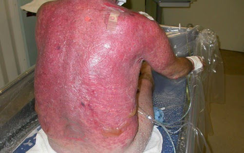
Figure 1: Patient with SJS / TEN - courtesy of Medscape

What is SJS/TEN  
These are severe mucocutaneous reactions most often triggered by medications, but can also be triggered by infectious processes (viral and bacterial).

The distinction between the two is based on severity.  SJS typically involves less than 10% of BSA, where as TEN involves >30% of BSA.  Both of these conditions often involve the mucous membranes (oral, ocular, or genital), in 90% of cases.

The overall mortality rate for SJS/TEN is approximately 30%, and the mortality is entirely dependent on the amount of BSA involved.


The most common drug culprits include: allopurinol, anti-seizure medications, antibiotics, and NSAIDs.

Clinical Syndrome
Cutaneous involvement - maculopapular rash on the trunk, and often sloughing of the skin "Nikolsky sign".
Mucous membrane involvement (90%)
Fever
Laboratory findings: elevation of LFTs, CBC abnormalities ,electrolyte disturbance.


Management
The cornerstone of management is supportive care (fluid management, wound care) and discontinuation of the underlying precipitant (i.e. allopurinol).  A monitored setting is required (ICU or Burn Unit), and consultations from ophthalmology and dermatology are necessary.

There is debate in the literature about the benefits of corticosteroids, IVIG, PLEX, as well as cyclosporine, so discussing the case with your local dermatologist is critical.

References
Association between HLA-B*58:01 allele and severe cutaneous adverse reactions with allopurinol in Han Chinese in Hong Kong. M.L.S. Chiu et al. British Journal of Dermatology. 2012.

Friday, November 28, 2014

NPH: Not Neil Patrick Harris, Normal Pressure Hydrocephalus

Today we discussed an interesting case of progressive dementia with marked gait abnormality.  The team described the gait abnormality as "magnetic".  The clinical course of this patient also included urinary incontinence, and ultimately an MRI confirmed enlarged ventricles, consistent with Normal Pressure Hydrocephalus.

Figure 1: Magnetic boots from Faceoff

What is NPH?

NPH is a rare disease characterized by the clinical triad of (gait disturbance, worsening cognitive function and urinary incontinence).  The gait classicaly in NPH is described as being "magnetic" as patients have great difficulty with taking a step, and lifting their feet off of the ground.  This gives the appearance that their shoes are magnetically attracted to the floor.

The pathophysiology of NPH is thought to be related to widespread dysfunction of CSF re-absorption at the level of the arachnoid granulation.  Over time, the ventriculomegaly and high levels of CSF causes edema, and microvascular ischemia leading to the neurological deficits.


Diagnosis:

The diagnosis is suspected if a patient has the clinical triad and neuroimaging showing ventriculomegaly and periventricular white matter edema.  The next best diagnostic test is a lumbar puncture to both confirm the normal CSF pressure, and also to demonstrate clinical improvement with the removal of >30 cc of CSF.  This is the so called "Fisher" test, where neurocognitive and gait assessments are done before and after the removal of large volumes of CSF, and if a patient has a signficiant clinical improvement than this would be consistent with the diagnosis of NPH.
Figure 2: Ventriculomegaly and white matter edema


Treatment:

The most widely utilized treatment in patients with confirmed NPH is the placement of a ventricular-peritoneal shunt (VP-shunt).  The reduces the amount of CSF in the ventricles and has demonstrated clinical improvement in many patients.  Other potential therapies include intermittent large volume lumbar punctures, and case reports also discuss the use of acetazolamide.

The Bottom Line:
NPH is one of the "reversible" causes of dementia, and internists need to have a high index of suspicion in order to make this diagnosis.  The diagnosis is also complicated by the fact that many patients with Alzheimer's dementia have urinary incontinence and also demonstrate ventriculomegaly related to underlying atrophy.  The "Fisher test", and having the persistence to perform a lumbar puncture and document clinical improvement is really the only way to make the diagnosis.

Reference: Normal Pressure Hydrocephalus: How often does the diagnosis hold water.  Klassen et al. Neurology 2011.

Wednesday, October 22, 2014

Hereditary Hemochromatosis - HH

Today we discussed an approach to a very classic internal medicine problem, a patient presenting with a new diagnosis of cirrhosis.  One of our former CMR's, recently inducted into the CMR hall of fame, Dr. Isaac Bogoch led the morning report!

Team 1 had a discussion about hereditary hemochromatosis (HH) as a potential cause for this presentation, which is a common genetic condition that is treatable if diagnosed early.

Genetic Features:

HH is an autosomal recessive iron-overload disorder associated with mutation of the HFE-gene on chromosome 6.  This results in a substitution of tyrosine for cysteine at position 282 of the HFE protein (C282Y).
http://www.marineinsight.com/

The original mutation is thought to be approximately 2000 years old, originating in northwestern Europe.  The defect is thought to provide women of child bearing age with some protection related to iron deficiency, and so propagated across northern Europe following the path of the Vikings.

The end result of this mutation in the HFE gene is high levels of iron being absorbed from the GI lumen, and dysregulation of iron storage and metabolism by both the liver and macrophages.  High levels of iron are released into the bloodstream and can deposit in various tissues leading to organ dysfunction.

The frequency of Heterozygotes amongst the caucasian population in the United States and Western Europe is approximately 10%

Ref: Pietrangelo. NEJM 2004. Hereditary Hemochromatosis.

Organs affected:

The main organs that are affected by iron deposition include the liver (leading to hepatits, and potentially cirrhosis), the heart (leading to cardiomyopathy), the pancreas (bronze diabetes), and various endocrine organs (the pituitary and testes).  HH is truly a multi-system disease.

Clinical Features:

The main clinical features include liver function abnormalities (75%), weakness (74%), skin pigmentation (70%), diabetes mellitus (48%), arthralgias (45%), and erectile dysfunction (45%)

Diagnosis:

A transferrin saturation > 45% is 95% specific for HH, and a serum ferritin level > 250 mcg/L is 85% specific in men, and 97% specific in women.  MRI or liver biopsy can also be helpful in making a diagnosis of iron overload states.

Ultimately, we have genetic testing for the HFE gene and other more uncommon genes (H63D) that can cause HH.

Women may have lower than normal ferritin levels because of menstruation, and so often the diagnosis of HH in women is not identified until after menopause.

Management:

The main treatment for HH is the ancient art of phlebotomy.  The target for phlebotomy is a ferritin usually less than 50.  It may take weekly phlebotomy (>500 mL of blood per week) in order to achieve this target.  Each 500 mL of blood contains approximately 250 mg of iron.  A patient with HH may have up to 10 grams (10 000 mg) of excess iron stores, so it may take many phlebotomies (1 per week, for a year) in order to achieve the target end point (ferritin <50 p="">
References:
Hereditary Hemochromatosis - A New Look at an Old Disease. Pietrangelo, A. 2004. New England Journal of Medicine.

UptoDate.

Wednesday, October 1, 2014

Lemierre Syndrome

A special thank you to Dr. Paul Bunce who did an amazing job and received an unprecedented ovation with morning report today.  Also, thank you to Team 1, Dr. Lindsay Melvin and Dr. Anthony Lott who presented a fascinating case!

Today we discussed a case of sepsis secondary to an acute oropharyngeal infection cause by the organism Fusobacterium necrophorum.

This prompted a discussion of the main syndrome associated with Fusobacterium necrophorum,   Lemierre Syndrome.

Some key learning points:

1.) Pharyngitis (sore throat) is the key presenting symptom - (80 - 87%) of cases
2.) There can be metastatic lesions (septic emboli) in the lung - 80% of cases
3.) Metronidazole or Clindamycin are the key antibiotics to cover Fusobacterium necrophorum
4.) Anticoagulation is controversial and is not the standard of care currently, but a consultation with hematology may be warranted.
5.) Early surgical intervention may be needed if the patient does not respond or improve with initial therapy.

Lemierre Syndrome:
The other name given to this condition is postanginal septicemia, which involves septic thrombophlebitis of the internal jugular vein, secondary to an acute oropharyngeal infection.  The syndrome was first characterized by Andre Lemierre (1875-1956) in 1936, who reported on dozens of cases from his own clinical experience.  He was a physician, and professor of microbiology and infectious diseases.  He published a description of 20 cases in The Lancet describing the clinical syndrome.

The key clinical features of this syndrome are that the initial source is usually within the pharynx (87% of cases), and IJV thrombus is present in approximately 70% of cases.  It can also cause metastatic lesions in the lung (80%) and joints (16% of cases).

The key symptoms and signs are sore throat (80%), fever (80%), and swollen or tender neck (52%).  Most of the diagnosis are made via cultures (70%), or radiology (IJ thrombus) - 15%.

The main laboratory findings are leukocytosis (75%) and thrombocytopenia (24%), and hyper bilirubinemia (32% of cases).
From: The Evolution of Lemierre Syndrome. Chirinos, et. al. Medicine. 2002

Most cases of Lemierre syndrome occur in younger patients (usually 16- 25 years of age).

The usual infectious agent in Lemierre syndrome is Fusobacterium necrophorum (82% of cases).  Other bacteria that can cause this syndrome include Bacteroides sp., and Group B and C Streptoccus.

This bacteria is a normal inhabitant of the oral cavity, and is a strict anaerobe, non-motile gram negative rod.  It has a characteristic appearance on gram stain (filaments, short rods, and coccoid elements).


Management:

Supportive Care:
A concern in any patient presenting with an oropharyngeal abscess is potential airway compromise, so stridor or any signs of respiratory distress should prompt a clinician to notify the ICU.  These patients can also become very sick, and may require ionotropic support.

Antimicrobials:
The initial management to a patient with suspected Lemierre syndrome includes antimicrobials with anaerobic coverage.  Either Metronidazole or Clindamycin are appropriate, with possibly additional coverage in the empiric setting for a polymicrobial infection.

Examples of empiric coverage: 
Cefazolin or Penicillin  G + Flagyl or Clindamycin

Vanco + Flagyl (if MRSA is suspected)

If the patient is septic, the "big guns" (Pip/Tazo, Meropenem) also provide excellent empiric coverage for this syndrome (gram positives, gram negatives, and anaerobic coverage).

Imaging:
Diagnostic Imaging: CT, MRI, or U/S to assess the neck and pharyngeal space, as well as imaging of the vessels is important.  Practically on call overnight a CT scan with contrast would be the imaging modality of choice.

Anticoagulation:   
The role of anticoagulation for an IJ thrombus in a patient with Lemierre syndrome is controversial. The outcome of most patients is very good (mortality is 6.4% in one case series).  There are no RCT's to clarify this question, but a discussion with Infectious Diseases and Hematology should be a part of the initial management plan.

Surgical Debridement / Source Control:  
If the patient has uncontrolled sepsis, or a more complication soft tissue infection (abscess, etc.)  Than a consultation with a surgical specialist is also warranted.

Reference:
The Evolution of Lemierre Syndrome.  Chirinos, J.A. et al. 2002. Medicine. 81: 458-65.

Lemierre Syndrome. Wright, W.F. et al. 2012. Southern Medical Journal. volume 105. Number 5.

Wednesday, September 24, 2014

The 4T's: Thymoma, Terrible Lymphoma, Teratoma, Thyroid cancer

Today in morning report we discussed an approach to a patient presenting with an anterior mediastinal mass.

Differential Diagnosis:  

The differential diagnosis for a mass in the anterior mediastinum includes the classic 4T's.

The most common primary tumours associated with this location are the following:

Thymoma - (30% of the time)
(Terrible) Lymphoma  - (20%)
Teratoma - and other germ cell tumours (18%)
Thyroid cancer (other carcinomas) - (13%)


Secondary tumours are more common in this region and include: lung, esophagus, and testicular cancers.

Clinical Presentation:

The presentation of patients with an anterior mediastinal mass is usually in the 3rd to 5th decade of life.
The main symptoms of a mass in this area include compressive symptoms (stridor, dysphagia, hoarseness) related to the mass impinging on surround structures.

Also, chest pain, cough, dyspnea are all common presenting complaints.

There can also be paraneoplastic phenomena related to the underlying malignancy, and we discussed how hypercalcemia can be associated with with lymphomas, and myasthenia gravis can be associated with thymomas.

The other important consideration is to not miss an aortic dissection in a patient presenting with chest pain and mediastinal fullness on a chest x ray.

Management:

There were a couple of important management considerations that we discussed.

1.) Assessing airway patency - if there is any evidence of stridor, or respiratory distress than a call to anesthesia or ICU to help intubate the patient is warranted.

2.) Ruling out Dissection - CT-angiogram to assess the mass and make sure this is not an aortic dissection.

3.) Obtaining a tissue sample - This can be performed via interventional radiology (IR) or respirology/thoracic surgery via EBUS (Endobronchial Ultrasound) guided biopsy.

Thymic epithelial tumours:

Thymomas have a predilection for females and usually present in the 5th and 6th decades of life.  Approximately 35% of patients with thymomas have myasthenia gravis, and 15% of patients with myasthenia gravis will have a thymoma.

Other associations with thymoma include pure red cell aplasia, hypogammaglobuminaemia, and SLE.

The bottom line as Dr. Mark Cheung would say, "When tumour is the rumour, tissue is the issue"


Reference: Imaging of anterior mediastinal tumours. Ching Ching Ong, Lynette L.S. Teo. 2012. Cancer Imaging. 12, 506-515.


Wednesday, September 10, 2014

SPICE: Inducible beta-lactamase in gram negative organisms


SPICE?

Today in Dr. HPK's morning report we discussed a very interesting case of sepsis secondary to a line infection.  In this case the blood cultures were positive for both Serratia and E.coli.  This brought up an interesting management question which we posed to the senior residents in the room:

What antimicrobial would you select empirically for such a patient?

There are a couple of questions to consider when faced with this particular management question:

1.) What is SPICE?


SPICE refers to a group of organisms that have a chromosomal beta-lactamase gene called ampC.  This beta-lactamase is active against beta-lactam antibiotics including cephalosporins, penicillin, and piperacillin/tazobactam.  The interesting point about this class of organisms is that they will produce the beta-lactamase enzyme only when stimulated by the presence of the antibiotic, hence the term "inducible".

The induced enzyme will be released into the periplasmic space and "eat up" the antibiotic, rendering it ineffective, resulting in treatment failure.

SPICE stands for: Serratia, Providencia, Indole-positive Proteus species, Citrobacter, and Enterobacter.  There are other gram negatives that have inducible beta-lactamase genes, but these are the most widely known species.

Serratia
Providencia
Indole-positive Proteus species
Citrobacter
Enterobacter

The various mechanisms that gram-negative bacteria can develop antibiotic resistance (NEJM, 2010)

2.) What antibiotics can I use to treat SPICE organisms?


The most important point to consider, is the avoidance of beta-lactam antibiotics.  

Beta-Lactam ring

Ceftriaxone - can you see the beta-lactam ring?

 Classes of antibiotics that do not induce the ampC gene include the following antibiotics:

TMP / SMX (Septra)  - oral / IV
Carbapenems (i.e. Meropenem) - IV
Aminoglycosides (i.e. gentamycin) - IV
Fluoroquinolones (i.e. moxifloxacin) - oral / IV

Comparison (by me): of the commonly used antibiotics to treat SPICE organisms

If a patient is septic (or for severe infections), than empiric therapy with an intravenous carbapenem is the ideal choice.

Consultation with an Infectious Diseases specialist and discussion with Antimicrobial Stewardship is also advisable in these cases.

Please check out the Antimicrobial Stewardship Website from UHN/MSH for more details, It is an amazing resource related to infections and antibiotics and can be accessed via mobile devices!

For further reading:
Hospital-Acquired Infections Due to Gram-Negative BacteriaAnton Y. Peleg, M.B., B.S., M.P.H. and David C. Hooper, M.D. The New England Journal of Medicine. 2010 May 13; 362(19): 1804–1813.

http://m.antimicrobialstewardship.com/clinical_summaries/index.php

Thursday, August 28, 2014

It's 3:00 AM and you want to do a thoracentesis?

Today in our Grand Morning Report, we discussed a common clinical dilemma that many Senior Medical Residents (SMR) face on call across the city:


This patient has a pleural effusion and it's 3:00 AM, should I perform a thoracentesis?


I would argue that there is really only one absolute, emergency indication to perform a thoracentesis on call (overnight) as an SMR covering the GIM service.

This being the old adage, "never let the sun set on an empyema".

If an SMR suspects empyema clinically, for example imaging confirms a loculated pleural effusion and the patient is febrile, than I believe the sun should not set, or in the case of our SMR's overnight, the sun should not rise before that pleural effusion is sampled and sent for culture.

This may require calling interventional radiology or thoracic surgery to provide assistance.

In my practical experience as an SMR at Sunnybrook Hospital, all that is needed overnight is an adequate sample to perform the necessary tests, mainly a sterile specimen for culture and gram stain, and to perform basic biochemistry (pH, cell count, protein, LDH).  This usually amounts to a 20 cc specimen, and no more.

Time... it is a precious commodity on call!


Practically on call, there is very little time to do a full therapeutic thoracentesis and drain off 1000 mL of fluid, so once the diagnostic sample is obtained, I would finalize the procedure and move on to the rest of your clinical duties.

It would not be an ideal scenario to be called to attend a Code Blue, while you are in the midst of draining off a large pleural effusion, and a needle is in the patient's back.

But... What if the patient is in respiratory distress and I feel that removing the fluid will alleviate their symptoms?


I would argue that the risks of performing a therapeutic thoracentesis overnight far outweigh the potential benefit.  If a patient is in such significant respiratory distress, that you want to insert a needle into their pleural cavity, they most likely will not have a significant clinical change from taking off 1000 mL of fluid.  

A unilateral pleural effusion is often not the only cause of the respiratory distress.  It usually takes an insult to both lungs to cause significant respiratory distress in most patients.  It is probably the presence of the underlying disease process such as pneumonia, congestive heart failure, malignancy, or pulmonary embolus that may be causing both the pleural effusion, and  the respiratory distress.

My suggestion in this circumstance would be to treat the underlying cause, such as giving lasix to a patient with CHF and a pleural effusion, provide supplemental oxygen (such as optiflow, or BiPAP), and if they are truly in respiratory distress, then these patients should be in a monitored setting (ICU or step down unit) as they will most likely require intubation or ventilatory support.  If it is patient discomfort that is the major problem, than providing symptom relief along with supplemental oxygen should be the way to go overnight.


What is the risk of performing a thoracentesis overnight?

Most of the literature points out that a bedside thoracentesis without the use of an ultrasound at the best of times carries a risk of pneumothorax between 4 - 30%, and approximately half of these patients will require insertion of a chest tube (Jones et. al, 2003).  Overnight, when an SMR has time constraints, and is fatigued this number is likely on the higher side.

Even with the use of ultrasound, the rates of pneumothorax were 2.5% among experienced interventional radiologists.

I love point of care ultrasound, but I am not an experienced interventional radiologist!

The Bottom Line:  


The last thing you want to do to a patient at 3:00 AM with significant respiratory distress is cause a pneumothorax as this may significantly worsen an already unstable situation.

The safe SMR knows when to subject a patient to a significant risk of harm, but more importantly knows when not to perform a procedure (especially overnight when there is very little support around).


If you're not ruling out empyema, don't stick a needle into the chest.  If the case is particularly challenging, call thoracic surgery or interventional radiology for assistance.  If the patient is unstable, call the ICU and if they are symptomatic, treat the symptoms and wait until the heavy cavalry arrives in the morning, and their is enough support around to safely perform the thoracentesis and deal with any serious complications.

A great reference on the rates of pneumothorax in the ultrasound era.  See reference below!

Ultrasound-Guided Thoracentesis: Is it a safer method? Jones, et. al. CHEST. 2003. 123: 418-423.


Wednesday, August 13, 2014

Thyroid Storm

Endocrine Emergency - Thyroid Storm

The theme for the week is management of the patient with critical illness, and today in Morning Report we discussed a case of thyroid storm.  This has been blogged before on Tangent's, but I wanted to focus more on the management of this medical emergency.

We also discussed the other thyroid related emergencies that might result in a patient being admitted to the ICU, mainly myxedema coma, which is the polar opposite of thyroid storm and hypokalemic periodic paralysis which classically presents as paralysis in male patients of Asian ancestry who are hyperthyroid.


Thyroid Storm:

In terms of thyroid storm, the Burch-Wartofsky scoring system is probably the most widely utilized clinical scoring system for making a diagnosis of thyorid storm.  The key clinical features in this scoring system include thermoregulatory dysfunction, CNS effects, GI-hepatic dysfunction, and cardiovascular dysfunction (including congestive heart failure and atrial fibrillation).  There is also often a precipitant (infection, trauma, surgery) that triggers the acute thyrotoxicosis.

Clinical pearl: thyrotoxicosis + hypotension
One clinical pearl that Dr. Silver mentioned was the presence of hypotension, in the context of a patient with symptoms and signs of thyrotoxicosis, should make a clinician very worried about true thyroid storm.

Management:
The management of thyroid storm includes the following:

1. Symptomatic treatment - In terms of managing symptoms beta blockers are the key component.  But other medications that can be helpful include acetaminophen 500-1000 mg PO q6h prn, which can be given to help manage the pyrexia.  Additionally, benzodiazepenes such as lorazepam can be given if the patient is very anxious and distressed.

Beta blockers such as propranolol can be given orally or intravenously if the patient is truly unstable.  The typical oral dose in thyroid storm is 40 - 80 mg PO q 4 hourly.  Beta-blockers assist with the psychomotor agitation, tachycardia, and also reduce the peripheral conversion of T4 to the more active T3.

2. Blocking the synthesis of new hormone - Thionamide (PTU)  - This class of medication, which mainly consists of Methimazole and PTU (propylthiouracil) blocks the synthesis of new thyroid hormone.  In the acute setting as Dr. Silver mentioned, PTU which has a rapid onset of action as compared with methimazole is the drug of choice.  If the patient is truly in thyroid storm than a loading dose of PTU may be given (1000 mg PO x 1 dose), then it is typically dosed TID in the 200 -400 mg range.

3. Reducing peripheral T4 conversion - Steroids - High dose steroids are often given for a variety of purposes.  The typical doses are 100 mg of hydrocortisone IV TID, or Prednisone 60 mg PO OD.  The duration is usually only for a couple of days.  The purpose of the steroids is to treat concomitant adrenal insufficiency that can be precipitated by the increased metabolism of endogenous cortisol as a result of the thyroid hormone excess.  Also, steroids decrease the peripheral conversion of T4 to T3.

4. Blocking the release of new thyroid hormone - Iodine - Once the patient has received a thionamide to block the synthesis of thyroid hormone, Iodine can be given as this will suppress the release of thyroid hormone from the thyroid gland.  Lugol's solution can be given, and as was mentioned today in morning report, it is usually only used for a short period of time (the initial 48-72 hours).
Other treatments that can be given if iodine is unavailable include Lithium, which also blocks the release of new thyroid hormone from the gland.

5. Treat the underlying precipitant - If is an infection, than appropriate antibiotics should be prescribed.

Lastly, a patient in true thyroid storm needs close monitoring (telemetry, frequent vital signs) and should be monitored in a high acuity setting such as a step-down unit or ICU.  These are also patients where your friendly neighbourhood Endocrinologist should be awoken in the middle of the night to assist in management.

Thanks to Dr. Robert Silver and team 3 for presenting this interesting case!

Reference:
Hampton, J. Thyroid Gland Disorder Emergencies. 2013.AACN Advanced Critical Care. Volume 24. number 3. pp. 325-332.


Wednesday, July 30, 2014

Viral infections and the kidney

Hepatitis C and the kidney

Thanks to Dr. David Frost and Team 1 for presenting an interesting case in morning report today:

We discussed hepatitis C, and the various mechanisms of acute kidney injury specific to the infection. 

Our case had a sub-acute course of 6 weeks duration in which the creatinine increased from a baseline of 69 to 677 on the day of presentation.

We also discussed the importance of the physical exam, making sure to search for findings suggestive of fluid overload, which may push you towards dialysis, and also looking for specific findings of cryoglobulinemia (palpable purpura).  In any patient with acute renal failure, searching for findings that might lead to urgent dialysis is of high priority and so listening for pericardial rubs, and looking for signs of uremic encephalopathy (asterixis, decreased LOC) is important.

Investigations:

The initial investigations showed a normocytic anemia.  There was a non-anion gap metabolic acidosis with a normal potassium.  The liver enzymes and function tests were normal.  The blood film, looking for findings of HHS or TTP, did not show any schistocytes.

The urinalysis, also known as the physical exam of the kidney, showed 3+ protein, trace blood, and the microscopy showed heme-granular casts.  The urine sodium was 24.  The abdominal ultrasound did not show any hydronephrosis or ascites, and the kidneys were normal in size.
 
Viral infections and the kidney: HIV, hepatitis B, and hepatitis C. Appel, G. Cleveland Clinic Journal of Medicine. Volume 74. May 2007.
Discussion:

This interesting case brought up a differential diagnosis that is not commonly encountered, but general internists should have some idea of how to approach it, mainly the various renal manifestations of viral infections.

Many viruses can cause renal impairment, most notably HIV (HIVAN), Hepatitis B -membranous glomerulonephritis (GN), and Hepatitis C which causes a membranoproliferative GN (MPGN).

Viruses can damage the kidney through different mechanisms including immune complex deposition such as in the case of cryoglobulinemia, through direct cytotoxic effects, and also as a result of the antiviral medications themselves.

Hepatitis C and the kidney:

In the case of Hepatitis C, possibilities included MPGN, cryoglobulinemia, adverse effects of the medications, and in patients with cirrhosis then hepatorenal syndrome.

The treatment of MPGN related to cryoglobulinemia associated with Hepatitis C often involves plasmapheresis (PLEX) to remove the cryoglobulins, immunomodulatory medications (steroids), and treatment for the Hepatitis C virus itself (interferon, ribavirin).

As Dr. Frost nicely pointed out, the time honoured approach of pre-renal, renal, post-renal with regards to the diagnosis and management of acute kidney injury almost never fails.  I must admit, I was taught this approach by Dr. J. Bargman as a 2nd year medical student.  I use it to this day, and I believe it will still be taught well off into the future.

Please see the excellent paper below, which discusses various viruses and their effects on the kidney:

Reference:

Viral infections and the kidney: HIV, hepatitis B, and hepatitis C. Appel, G. Cleveland Clinic Journal of Medicine. Volume 74. May 2007.

Wednesday, July 16, 2014

Endocrine Morning Report - Euglycemic DKA in pregnancy

A devastating complication of pregnancy in type I diabetics

A special thank you to Dr. Robert Silver for leading our discussion of the case.

           Today we discussed an interesting case of a 29 year old G1P0 female who presented at 32 weeks gestation with a chief complaint of shortness of breath.

On her third day of admission in hospital, she developed worsening of her symptoms and her respiratory rate increased to 40 breaths per minute, with a normal oxygen saturation (98%) on room air.

Her capillary blood glucose registered a blood sugar of 10 mmol/L, and it was only after examining her arterial blood gas that a diagnosis of diabetic ketoacidosis (DKA) could be established.

Her arterial blood gas (ABG) showed a pH of 7.29 / pCO2 of 14 / pO2 of 120 / HCO3 of 6.  Her serum electrolytes showed an anion gap of 31, and alarmingly a serum potassium of 4.7 (in the context of insulin deficiency, this is worrisome).

The appropriate intervention, being transfer to the intensive care unit, potassium replacement via a central line, and aggressive fluid resuscitation were all initiated. She was started on an insulin infusion as well as IV D10W to maintain her blood sugar, and within 24 hours she was completely stable and back to her normal state of health.  

Table 1: The American Diabetes Association diagnostic criteria for DKA: triad of hyperglycemia, anion gap metabolic acidosis, and ketonemia.


Why do pregnant patient's decompensate into DKA at a lower glucose?

           There are a number of cases reported in the literature of euglycemic DKA in pregnant patients, and it is thought to be a rare but devastating complication of pregnancy in type 1 diabetics.  The frequency of occurrence is approximately 1% of all DKA's in pregnant patients (Guo, 2008).  It carries a high fetal mortality rate (30- 90%) and is often a missed diagnosis because of the falsely reassuring glucose readings (Guo, 2008).

Maternal Changes:
The pathophysiology of this process is  interesting.  The normal physiology of pregnancy results in a decrease in insulin sensitivity, and this is thought to be related to increased lipolysis and ketogenesis (Chico et. al, 2008).  Also, pregnant women have a baseline respiratory alkalosis due to pregnancy induced hyperventilation.  This results in a decreased serum bicarbonate and reduced buffering capacity (Chico et. al, 2008).

These normal physiologic changes reduce the body's ability to cope with acidosis, whether it be sepsis or DKA mediated.

Placental-Fetal Changes:
Furthermore, the growing placenta and fetus utilize large quantities of glucose and produce factors such as placental lactogen, progesterone and cortisol that further increase insulin resistance, especially towards the later stages of pregnancy.

The end product of both these maternal-fetal changes is that pregnant patients with T1DM are at an increased risk of developing DKA.  They also have a lower serum glucose threshold at which DKA will occur compared to non-pregnant women with T1DM (Guo, 2008).


The average glucose of pregnant patients with DKA was 16 mmol/L compared with 27 mmol/L in non-pregnant patients (Guo, 2008).

Take home message: In pregnant women with T1DM, a normal appearing glucose does not adequately rule out DKA and can have devastating consequences for both mother and the fetus.


Please see the video below about Dr. Ho Ping Kong and his best selling book "The Art of Medicine".

References:
Guo, R.X., Yang, L.Z., Li, L.X., Zhao, X.P. Diabetic Ketoacidosis in pregnancy tends to occur at lower blood glucose levels: case-control study and a case report of euglycemic diabetic ketoacidosis in pregnancy. Journal of Obstetrical Gynaecology Research. 34(3):324-330. 2008.


Chico, M., Levine, S.N., Lewis, D.F. Normoglycemic diabetic ketoacidosis in pregnancy. Journal of Perinatology. 28(4): 310-2. 2008.


Thursday, July 3, 2014

Welcome to the 2014-2015 Academic Year! Emergency Lecture Series - Upper GI Bleed

Firstly, I just wanted to thank the outgoing CMR's for all of their hardwork and dedication over the past year.  I look forward to continued friendship and working with you guys into the future.

Now we shall get to business...

Our first blog today is about the Emergency Management of an Upper GI Bleed:

We discussed at lunch rounds today that there are 5 essential questions that need to be answered by the Internist in the emergency management of any patient with an upper GI bleed (UGI bleed).

Question #1:  Is this truly an upper GI bleed, or is it something else (hemoptysis, a lower GI bleed):
From the JAMA rational clinical exam series, useful historical features to help you answer this question include whether they've had a previous UGI bleed (LR+ of 6.2), if their is a history of melena (LR+ 5.5), cirrhosis (LR+ 3.1) and if they have been taking any anti-coagulation (mainly warfarin, but can likely be extrapolated to the NOAC's) - LR+2.3.

The most useful feature on physical exam to determine if the patient has an UGI bleed would be the presence of melena on DRE (LR+ 25).

Question #2: How severe is this UGI bleed?

Again, from the same paper, key historical features that predict a more severe UGI bleed include a history of malignancy or cirrhosis (LR + 3.7), and a history of syncope (LR+ 3.0).  On the physical exam, abnormal vital signs, mainly tachycardia (HR>100) has a (LR+4.9) for a severe UGI bleed.  The presence of an orthostatic drop in the blood pressure also is suggestive of a more severe UGI bleed (LR+ 2.8).

Luckily, in the 21st century a clinician does not have to memorize these numbers and they have been nicely integrated into a clinical score (Glasgow-Blatchford Score) - which can be accessed at the following link

Question #3: Does this patient have a variceal bleed?

To assist with answering this question from a clinical perspective, the JAMA series again proves useful.  Mainly, once you have determined the patient is having an UGI bleed, the next step is to search for signs suggestive of cirrhosis.  If any features of cirrhosis are present, then the safest approach would be to presume the bleed is variceal in origin.  Unfortunately in determining whether a patient has cirrhosis, history does not prove very useful.  The physical exam and baseline laboratory investigations are however, very useful.  The presence of distended abdominal veins "caput medusae" is particularly helpful in ruling in cirrhosis (LR+ 11).  Other helpful clues include encephalopathy (LR+10), ascites (LR+ 7.2) and jaundice (LR+ 3.8).

From a laboratory perspective, the most helpful clues to making a diagnosis of cirrhosis include thrombocytopenia (LR+ 9.8) if the platelets are less than 110, a prolonged INR (LR+ 5.0), and a low albumin (LR+ 4.4).

Again, if one suspects a variceal bleed, it should be treated as such.

Question #4: Does this patient need ICU or urgent endoscopy?

To answer this question, if there is hemodynamic compromise such as shock (elevated lactate, etc.) or signs of organ hypoperfusion (demand ischemia), the threshold for calling help is quite low.  For severe UGI bleeds (based on Glasgow-Blatchford score), or those with high risk features (hypotension, tachycardia), urgent endoscopy and close monitoring is essential.  If there is the suspicion of a variceal bleed, then ICU and GI should be notified in most cases immediately to help expedite endoscopy.

Question #5: Is the patient on an anticoagulant, and what is the indication?

This should essentially prompt the internist to consider the risks and benefits of reversing the anticoagulation.  These decisions are on a case-by-case basis and almost always should include input from gastroenterology, cardiology (in the case of mechanical valves), and hematology (in the cases of DVT/PE).  With the newer anti-coagulants, a phone call to the blood bank and ICU is also necessary as currently their exists no anti-dote or reversal agent on the market.

Those are the key questions to consider in the emergency management of an UGI bleed from the intern, or internist's perspective.  Please see the below references for further details on the management of an UGI bleed.

That was a long tangent... Welcome to 2014-2015
(Thanks Lee)

Management of Acute Bleeding from a Peptic Ulcer. Gralnek, et. al. NEJM. 2008
Does this patient have a severe Upper Gastrointestinal Bleed. Srygley, et. al. JAMA. 2012.
Does this patient with liver disease have cirrhosis. Udell, et. al. JAMA. 2012

Thursday, April 24, 2014

Renal tubular acidosis

Today we discussed a case of acidosis. A young patient presenting with a significant metabolic acidosis of mixed origin (both anion gap and non-anion gap). This was determined by carefully examining the patient electrolytes and blood gas results, identifying the primary acid-base abnormality and calculating what is called the delta-delta.

This refers to the difference between the calculated and expected anion gap and the difference in the bicarbonate from normal serum values. When the bicarbonate value is lower than what would be expected from the anion gap difference (a 1:1 change with respect to AG and bicarbonate) and additional non-anion gap acidosis is identified. When the bicarbonate is higher than expected, a concomitant metabolic alkalosis is diagnosed.

Anion gap acidosis is a commonly encountered scenario, and there are widespread mnemonics to help you consider the differential diagnosis (ex. MUDPILES). Non-anion gap metabolic acidosis seem to be less commonly discussed, but are worth reviewing. The differential diagnosis includes:

Resuscitation with hyperchloremic solution (NS)/hyperalimentation
Medications: acetazolimide
Renal tubular acidosis
Diarrhea
Anatomic fistulas: uretoenteric fistuls and pancreaticoduodeneal fistulas

The patient today was known to have chronic metabolic acidosis, raising a RTA as a likely cause. These can be divided anatomically into four categories:

a) proximal RTA (type 2) - here there is an abnormality resulting in the reabsorption of bicarbonate. Patient tend to have mild to moderate acidosis with a relative ability to excrete acid in the form of NH4Cl-, and acidify the urine.

Proximal RTA can be thought of as primary and secondary. There are congenital syndromes associated with this, some of which have associated visual and cognitive deficits. One must look for evidence of the Fanconi's syndrome, where in addition to inability reabsorb bicarbonate, glucose, phosphate and potassium are lost in the urine. When present, paraproteinemia (multiple myeloma), should be considered, given the light chains can block re-absorption of electrolytes in the tubules. Drugs and other metabolic and congenital diseases may also present with this and should be considered (See attached article for details). Other things to watch for include bone disease and renal stones with increased loss of electrolytes in the urine.

b)distal RTA (type 1) - results from the inability to excrete protons at the level of the distal tubules. This results in severe acidosis and inability to acidify the urine. Again, this can be a result of congenital disease (often associated with sensorineural hearing loss because of shared gene expression in the cochlea and the kidney) and acquired. A detailed history for connective tissue and autoimmune disease should be performed, with rheumatoid arthritis and Sjogrens syndrome as common entities identified.

c) Mixed proximal/distal (type 3) - these patients have features of both distal and proximal RTA. Rare mutations in the carbonic anhydrase gene have been attributed to mixed RTA. Other clinical features include osteopetrosis, cerebral calcification and mental retardation.

d) Hyperkalemia RTA (type 4) - is due to impaired ammoniagenesis. Most common causes include hyporeninemic states as a result of primary renal disease (diabetic nephropathy) or medications that cause hypoaldosternonism (spironolactone). Congenital syndromes do exist causing pseudohypoaldosteronism however these are uncommon.

For additional details regarding the diagnosis, investigations and treatment of RTA please see the below review article which provides a good overview.

RTA review

Friday, April 11, 2014

Therapeutic hypothermia

Today we discussed a complicated case of multifocal sepsis in a patient with hypoxic ischemic injury following cardiac arrest. This provides an opportunity to discuss neurologic recovery following cardiac arrest, the role for cooling as part of post-resuscitation care and examining for coma/brain death.

Cardiopulmonary resuscitation has been around since the 1950's, but its practices weren't widely promoted until the 1970's when the promotion of CPR began in the public sector as well. Since then, it has involved from chest compressions and breathing support, to add defibrillation and consideration of advanced life support techniques. In 2012, a Cochrane review looked at several studies from the previous decades at the benefit of post cardiac arrest therapeutic hypothermia, finding it to be beneficial in terms if patient in hospital neurologic recovery and overall survival. This advantages were seen without any adverse effects. This supported the current guideline recommendations, which include therapeutic hypothermia after cardiac arrest.

Although this is not as relevant for residents rotating through internal medicine, many will at some point complete ICU, CCU, cardiology and emergency medicine rotations, where this practice is important. The basics of therapeutic hypothermia should be known, and include the following:

1. Indications
2. Implementation
3. Rewarming
4. Contraindications

Patients with cardiac arrest that do not have a purposeful movement and are not following commands after return of spontaneous circulation should be considered for therapeutic cooling. There are few contraindications. The things to recognize are the risks of coagulopathy in the bleeding patient and electrolytes shifts (particularly potassium) that can occur with cooling and rewarming. Cooling can be accomplished by many methods, including external cooling blankets, ice packs, cold IV fluids (4 degrees) or internal catheter devices. Which to choose depends on the expertise of the centre and comfort of the physician. Cold IV fluids can rapidly degrees core temperature and run the risk of pushing the patient into pulmonary edema, which should be considered if this method is chosen. Targets for therapeutic cooling are somewhat controversial. A target of 33 degrees is likely reasonable in the first 24 hours, with gradual rewarming until 48 hours is reached. Whether or not cooling should be performed for over 48 hours is unclear. A recent paper in the NEJM, found no difference in outcomes when targeting a core temperature 33 vs 36 degrees. See below for a link to the original article.

After the patient is normothermic, an assessment of neurologic status is needed to try and predict neurologic recovery. Predicting neurologic prognosis after cardiac arrest has the most literature, as compared to neurologic injury from other causes. Several systematic reviews have been performed, and found that the useful predictors include:

Absent pupillary response/absent corneal response at 72h
Absent extensor motor response at 72h

One of the original articles is linke below from Neurology.

Predicting neurologic outcome after cardiac arrest
NEJM cooling after cardiac arrest

Thursday, January 9, 2014

Flash pulmonary edema

Acute onset shortness of breath is a common reason for referral to GIM and congestive heart failure is in the top three admission diagnoses in most hospitals. Flash pulmonary edema is a rapid development of pulmonary congestion, usually secondary to the development of increased left ventricular diastolic pressure. It can present quite dramatically, as the name suggests, having impressive pulmonary infiltrates, dyspnea and hypoxemia.

Risk factors for flash pulmonary edema are similar to other risk factors for heart failure, including:

HTN
ischemic heart disease
valvular heart disease
diastolic dysfunction
bilateral renal arterty stenosis

The diastolic dysfunction that leads to high pressures may be suggested on echocardiography. First, we should review the phases of diastole, which include:

Isovolumetric phase
early atrial empyting
stasis phase
atrial contraction

On echo, the early (E) to atrial contraction (A) phase should be relatively high, where E >75% of the interval. As diastolic dysfunction develops this ratio will decrease <1 .="" a="" additional="" and="" can="" changes="" eventually="" occur="" over="" pseudonormalization="" ratio="" see="" time="" where="" you="">2 suggesting restricted filling. 
This can be seen in patients with chronic diastolic dysfunction.

Mitral regurgitation is another cause of flash pulmonary edema. If there is ischemia to the papillary muscles they can rupture or become dysfunctional. This would be evident by a loud regurgitant holosystolic murmur best heard at the apex. Ischemia can be related to anterolateral papillary muscle dysfunction or posteromedial dysfunction. The anterolateral muscle is supplied the LAD and Cx artery, while the posteromedial tends to be only supplied by the posterior descending artery (PDA). As a result the collateral circulation of the anterolateral muscle makes it less susceptible to rupture. The PDA can stem from several possible arteries, which determines the dominance of the coronary circulation:

A "right dominant" circulation occurs when the PDA stems from the RCA.
A " left dominant" circulation occurs when the PDA stems from the Cx.
A "codominant"circulation occurs when the PDA stems from the Cx and RCA.

In the setting of acute MR from ischemia, cardiovascular surgery may be required. Surgery should also be considered in patient with severe MR (regurg fraction >50%, orifice >0.4cm, volume of regurgitation >60%

Approximately 70% of the population is right dominant, where only 10% are left dominant.

Renal artery stenosis, whether artherosclerotic or fibromuscular dysplasia can result in flash pulmonary edema (formerly known as pickering syndrome). Overall it may cause up to 5% of HTN in adults. Patient with refractory HTN or flash pulmonary edema may be considered for renal artery stenting, however there another recent trial that suggested this intervention is no better than medical therapy alone. Think about RAS in the following settings:

1. Worsening kidney function of 30% in started and ACEi
2. Systolic/Diastolic renal bruit
3. Onset HTN after 55
4. Flash pulmonary edema
5. Asymmetry in renal size >1.5 cm (see previous RAS blog post)

Here is the latest NEJM article on RAS stenting efficacy

NEJM RAS stent vs medical managment


Friday, January 3, 2014

Pyogenic liver abscess

Discussion today surrounded an atypical presentation of a pyogenic liver abscess. Typically, this condition will present with fever, sweats, abdominal pain (usually RUQ) and occasionally symptoms of systemic infection. Todays case was a bit strange, given the only consistent features were constitutional symptoms, and atypical chest and abdominal pain were misleading. As a result, one must have a high index of suspicion for a liver abscess with fever of unknown origin (FUO). Following negative preliminary testing in the evaluation of FUO abdominal imaging should be considered, especially when there are abnormal liver enzymes. This approach led to a diagnosis in 19% of cases where first line tests were unrevealing.

Pyogenic liver abscess is the most common visceral location for focal infection. That being said, its really not that common, where some studies quote 2-3 cases per 100,000. Most studies on epidemiology of liver abscess have been performed in South East Asia, and may even be more uncommon in our population.

These infections can develop through several mechanisms:

1. Hematologic bacteremia leading to seeding
2. Biliary obstruction and reflux of bacteria into liver parenchyma
3. Direct invasion through adjacent structures (gastric perforation) or contact through bowel leakage/perforation

Most often these infections involve the right side of the liver given the blood supply tends to be larger for this lobe.

Many micro-organisms can cause liver abscess formation. Some of these include:

Strep. anginosis, staph. aureus and strep pyogenes are amongst the most common pathogens involved. Klebsiella sp., as in our patient, which is a gram negative abscess forming bacteria is also well described. There was a large case series from Taiwan which found that Klebsiella was a common cause of liver abscess (nearly 70%), most often developed in the community, and was associated with DMII and hyperglycemia.

The treatment of these patients is antibiotic therapy, with a strong focus on source control. Abscess drainage can be both therapeutic and diagnostic and is a required part of therapy unless the abscess are too small for drainage. Antibiotic therapy should be targeted towards the identified infection, and duration consist of at least 4 weeks of IV antimicrobials.

One case series of Klebsiella liver abscess patients found an overall mortality rate of nearly 10%, and a relapse rate of 4%. Sepsis was the cause of death in all of those who died. This organism can be aggressive and patients should be evaluated for additional metastatic sites of infection.