Tuesday, July 31, 2012

Today we talked about a case of acute hypoxia in an elderly gentlemen with a history of dysphagia, stroke, seizures and dementia.We covered the differential for acute hypoxia and then identified that the most likely cause in our patient was aspiration pneumonia, with pulmonary embolus on the differential.

1) Causes of Acute Hypoxia: The differential for hypoxia is extensive, but the differential for sudden onset severe hypoxia is not as long.

  • Pulmonary embolus
  • Pneumothorax
  • Pulmonary edema (cardiogenic/non-cardiogenic)
  • Pulmonary hemmorhage
  • Aspiration pneumonia
  • Bacterial pneumonia
  • Mucous plugging
2) Aspiration: Defined as misdirection of oropharyngeal or gastric contents into the larynx and lower respiratory tract. There are two entities: Aspiration pneumonitis and pneumonia. Aspiration pneumonitis can lead to ARDS and carries a high mortality (up to 12%). It can present with fever and hypoxia. Treatment is supportive. Use of glucocorticoids is controversial and animal studies have shown variable results.

Half of healthy adults aspirate during their sleep, but are protected from developing aspiration pneumonia by the following mechanisms: low virulent bacterial load in orophargyngeal/gastric contents, active ciliary transport, normal cough reflex, cellular and humoral immune system to help clear infection.

  • Risk factors for aspiration include
    • Decreased LOC caused by drugs or toxins (i.e. EtOH)
    • Stroke/neurologic injury causing dysphagia/impaired cough reflex
    • Seizure disorder
    • Degenerative CNS disease: dementia, Parkinson's, ALS
    • Dysphagia: esophageal stricutre, diverticulum etc.
    • Recent anesthetic
    • ETT/Trach/NG tube
    • Frequent reflux/vomitting/gastric outlet obstruction
  • Risk factors for developing aspiration pneumonia:
    • Poor dentition
    • Treatment with PPI
    • Underlying lung disease
    • Immunosuppression/decreased cellular immunity
    • Colonization of oropharynx with s. aureus, GNB (Klebsiella pneumonia, Escherichia coli)
  • Common bacterial pathogens:
    • Oral anaerobes: Fusobacterium, Bacteroids, Peptostreptococcus
    • Gram negative bacilli: Klebsiella, E. coli
    • S. aureus
  • Management:
    • Swallowing assessment: by SLP +/- Video Fluoroscopic Swallowing Study (VFSS)
    • Dysphagia diet
    • Antibiotics
      • When anaerobic bacteria is the most likely pathogen: consider Clindamycin
      • Other regimens that also cover CAP: 
        • Amoxicillin-Clavulanate
        • Moxifloxacin
        • Flagyl plus Amoxicillin or Penicillin
      • For nosocomial pneumonia and pt is unwell and coverage for both aerobic and anaerobic bacteria is important: 
        • Pipericillin-tazobactam
        • Carbapenems
    • Management of dysphagia:
      • Studies have looked at using dopamine agonists and ACEi to increase substance P, which enhances swallowing and the cough refelx.
      • Percutaneous feeding/PEG tube has not been shown to increase survival, quality of life or decreased aspiration events
The following is a systematic review of the evaluation and management of dysphagia due to dementia in the elderly: 
http://bf4dv7zn3u.search.serialssolutions.com.myaccess.library.utoronto.ca/OpenURL_local?sid=Entrez:PubMed&id=pmid:22608838

Friday, July 20, 2012


Today Dr. Okrainec took us through a very interesting case of an unstable patient with an UGIB.
The medical students, R1s and R2s did a great job of stabilizing the patient and eventually saving his life! Here are some key points that came out of this morning's session.

1) Approach to the unstable patient:

  • Always remember your ABC's
    • Be cognisant of changes from a patient's baseline, a drop of more than 20mmHg from the baseline BP is significant, even if the absolute value is not in and of itself alarming.
    • Does the patient look sick? Sweating, clammy, cold extremities
    • Always reassess vitals when there is a change in pt's status and Q2-5 minutes
    • Call for help: when needed you can call a code blue to get a 1) monitor 2) ICU nurse 3) RT/anesthesia 4) crash cart
    • Gather as much information as possible: delegate a member on your team to collect information such as: meds, last blood work, past medical history, major things to look at are last creatinine, INR, hgb, wbc
    • Do a focused physical exam
    • Always plan ahead: Check that you have adequate IV access, inform the ICU of a sick patient, will you likely need blood (do you have an up to date group&screen in the lab?)
2) Approach to UGIB
  • ABCs
    • Start with getting 2 large bore (16-18G) IVs 
    • Start with fluid resuscitation
    • Get blood: Uncross matched if pt is hemodynamically unstable and low likelihood of having alloantibodies.
  • Initial Management:
    • Start Pantoloc 80mg IV bolus followed by 8mg/hr infusion
    • If hx or risk factors for liver disease and varices start octreotide
    • Consider NG tube, but careful if pt has known esophageal varices
    • Optimize clot formation:
      • Reverse INR: Octreotide + Vit K
      • Platelets: should be above 50
      • Uremia: can give DDAVP to help platelet function
  • Definitive management:
    • Call GI: for early endoscopic treatment
    • Call ICU: for possible intubation
    • If pt has liver disease and ascites consider prophylaxis for subacute bacterial peritonitis (SBP)


Thursday, July 19, 2012

Today we had a great discussion on bread and butter Internal Medicine cases from the night before. Here is a tasting menu of the issues that came up in this morning's "Grand Morning Report"

1) Proton Pump Inhibitors - potential side effects

  • Proton pump inhibitors have revolutionized the treatment of UGIB. An acidic environment inhibits   platelet aggregation and promotes fibrinolysis of formed clot by the enzyme pepsin. Bringing the gastric pH to above 6 is thought to promote clot formation. Studies have shown that treatment with a PPI signficantly reduces recurrent bleeding secondary to PUD. 
  • PPIs are often started for various reasons, i.e. to promote healing of peptic and duodenal ulcers, symptom relief from GERD etc. They are very effective, however many forget to reassess the need to continue on PPI therapy and patients may continue them indefinitely. This has led to long term complications of PPI use.
  • Nutritional:
    • Protein bound dietary Vitamin B12 requires acid and pepsin for its initial absorption. Vitamin B12 then binds to haptocorrin, found in the saliva. It is then liberated in the higher pH environment of the duodenum, where it binds to intrinsic factor and is absorbed in the terminal ileum.This may be more important for people who have low Vit B12 intake (i.e. Vegans) or the elderly. Therefore it may be reasonable to measure Vit B12 levels in these populations of people who are on PPIs.
    • There is a theoretical possibility of iron deficiency, as gastric acid is needed to reduce non heme iron (Ferric iron) to the more soluble Ferrous iron.
    • Osteoporosis: Gastric acid secretion is important for dietary calcium absorption.
  • Infections:
    • Pneumonia: By changing the gastric flora and seeding of the lungs from the upper alimentary tract.
    • C. difficile and other enteric infections: Also due to changes in the gastric flora 
  • Cancer:
    • Theoretically the hypergastrinemia resulting from chronic acid suppression can lead to hyperplasia of the gastric enterochromaffin-like cells and lead to gastric carcinoid tumours. Gastrin is also trophic for colonic mucosa, and may also lead to colorectal carcinoma.
2) Treatment of bacteriuria in the Elderly
  • There is no evidence for treatment of asymptomatic bacteriuria in the elderly. The only cases where asymptomatic bacteriuria should be treated is in pregnant patients and prior to urologic procedures.
  • Bottom line, a positive urinalysis in an obtunded elderly patient does not mean the patient has urosepsis! Look for other signs of infection: Fever, white blood cell count, imaging findings of pyelonephritis, lower urinary tract symptoms.
3) When to suspect Legionella?
  • Fever >39C
  • Neurologic findings, confusion
  • Gastrointestinal findings: Nausea/Vomiting/Diarrhea
  • Transaminitis
  • Leukocytosis, thrombocytopenia
  • Hematuria
  • Hyponatremia
  • Failure to respond to beta-lactams or aminoglycocide
  • CXR: can show anything from lobar consolidation to interstitial pulmonary inflitrates

Friday, July 13, 2012

Paroxysmal Nocturnal Hemoglobinuria


Today we talked about Paroxysmal Nocturnal Hemoglobinuria, an interesting cause of intravascular hemolysis and thrombosis.

1) Pathogenesis:

  • Defect in the PIG-A gene that encodes the cell membrane anchor glycophosphatidylinositol (GPI). Glycophosphatidylinositol is necessary for binding proteins to the RBC membrane.
  • Defect in the GPI anchor leads to the absence of GPI linked proteins. Two important proteins that are missing include CD55 and CD59, which are involved in the down regulation of the complement system.
  • The complement system is part of our innate immunity and is composed of a cascade of enzymatic reactions that culminate in the creation of a membrane attack complex (MAC) that results in cell lysis and destruction. There are two forms of the complement system, the classical pathway that is initiated by IgG/IgM antibodies and the surveillance system that does not require antibodies to become activated. It is this latter system that affects RBCs. 
  • CD55 and CD59 act to accelerate decay of the complement system and protect against lysis, respectively, thereby preventing the formation of the deadly MAC. Patients with PNH who lack CD55/CD59 suffer from episodic intravascular hemolysis, the severity of which depends on the degree of absence of GPI linked proteins.
    • PNH III: complete absence
    • PNH II: partial absence
    • PNH I: normal

2) Symptoms
  • Hemolysis: Episodic hemolysis, the severity of which depends on the degree of expression of CD55/59. The hemolysis is intravascular and therefore characterized by hemoglobinuria, hemoglobinemia and hemosiderin in the urine. Hemolysis in PNH can happen at any time, not necessary at night as the name implies. However, it is thought that hemolysis increases at night due to intestinal absorption of lipopolysaccharides that enhance the complement system. 
  • Nitric Oxide sequestration: Free hemoglobin is a NO scavenger. Nitric Oxide is important in relaxing smooth muscle, therefore depletion of NO as a result of hemolysis results in smooth muscle dysfunction. This manifests as esophageal dysmotility with dysphagia and abdominal cramping.
  • Renal dysfunction: Acute hemolysis results in hemoglobinuria that can cause acute kidney injury. Also over time, chronic renal dysfunction occurs due to hemosiderin deposition.
  • Prothrombotic state: Patient with PNH have a higher propensity for both venous and, to a lesser extent, arterial thrombosis. The etiology is not well known but thought to be secondary to an abnormal  line of platelets. Patients present with thrombosis of hepatic veins, intra-abdominal veins and even cerebral veins.
  • Diminished hematopoiesis: May progress to aplastic anemia
  • Hematologic malignancies: May lead to myelodysplastic syndrome and acute leukemia.
3) Treatment
  • The treatment of PNH has been revolutionized by the introduction of eculizumab, a humanized monoclonal antibody against C5. This stops terminal complement activation. See this link to the TRIUMPH study a double blind randomized multicenter control study looking at use of eculizumab compared to placebo for PNH. TRIUMPH
  • Supportive therapy includes intermittent transfusions as needed and iron/folate supplementation. Be careful of patients developing iron overload due to repeated blood transfusions!
  • Anticoagulation is initiated for those with a history of thrombosis. Prophylactic anticoagulation may also be used in those with a large percentage of abnormal granulocyte clones.


Wednesday, July 11, 2012

Today we discussed an interesting case of a 21 year old male with a non-healing ulcer on his shin. Interestingly he had been recently diagnosed with ulcerative colitis. While, the diagnosis of pyoderma gangrenosum seemed most obvious, the biopsy in fact showed evidence of leukocytoclastic vasculitis and the patient was found to have high peripheral eosinophils and was P-ANCA positive. Talk about a curve ball!

Here are some interesting questions that arose from our discussion:

1) Differential Diagnosis of ulcerative lesion:
Refer to the following NEJM article for an interesting discussion of skin ulcers misdiagnosed as pyoderma gangrenosum. This article gives a great differential diagnosis for ulcerative skin lesions. The key is to have a broad differential and to do a biopsy! The treatment for pyoderma gangrenosum is not benign (steroids and other immunosuppressants) so be sure of the diagnosis before you treat.

2) Leukocytoclastic vasculitis is not a diagnosis in and of itself but a pathologic term describing neutrophilic small vessel vasculitis. It is an indication of small-vessel vasculitis. This can occur in the following settings:

  • ANCA associated small vessel vasculitis:  
    • Granulomatosis with polyangiitis (formerly known as Wegener's vasculitis)
    • Churg-Strauss vasculitis
    • Microscopic Polyangiitis
  • Immune Complex associated small vessel vasculitis:
    • Hepatitis C with cryoglobulinemia
    • Henoch-Schonlein pupura (HSP)
    • Connective tissue disease-associated vasculitis:
      • RA
      • SLE
      • Sjogren's Syndrome
    • Endocarditis
  • Hypersensitivy vasculitis:
    • Penicillin, ASA, amphetamines, thiazides
  • Viral infections:
    • Strep throat
    • bacterial endocarditis
    • TB
    • hepatitis
    • Staphylococcal infections
    • Foreign proteins (serum sickness)
3) Differential for non-vasculitic P-ANCA positivity:

  • Infectious: HIV, mycobacterial infections, severe pneumonia, bacterial endocarditis
  • Chronic: Rheumaotid arthritis, IBD, Sweet's syndrome, eosinophilia-myalgia syndrome, Goodpasture's syndrome
  • Neoplasms: Atrial myxoma, small cell lung cancer, NHL, myelodysplasia, colon carcinoma


4) Differential for Eosinophillia:
  • Infectious: Parasitic infection, fungal infection (aspergillosis)
  • Hematologic/neoplastic: Hypereosinophilic syndromes, leukemia, lymphoma
  • Allergic disorders: atopic dermatitis, asthma, rhinitis, medications
  • Rheumatic disease: Churg Strauss
  • Miscellaneous: Adrenal insufficiency, cholesterol embolization


Tuesday, July 10, 2012

This morning we discussed an interesting case of an 84 year old male who presented with falls and change in mental status. Dr Nadjafi took us through an approach to delirium (see blog from July 5th for approach to delirium). After thorough investigations it was discovered that the patient's Calcium was 3.98mmol/L! Here is a quick approach to hypercalcemia:

1) Diagnosis:
- Serum Calcium: 40-50% of calcium in serum is bound to protein (mainly albumin). It is the ionized (or "free") calcium that is physiologically important. Therefore, hypo- or hyper-albuminemia can affect the serum ionized calcium. The following calculation helps you determine ionized calcium from measured calcium.

Ionized Calcium = measured Ca + 0.2(40-measured albumin)

- Degree of hypercalcemia: Mild hypercalcemia (<2.75mmol/L) usually seen in hyperparathyroidism. Severe hypercalcemia (>3.25mmol/L) usually indicates malignancy associated hypercalcemia.

2) Symptoms:
 
Neuropsychiatric
Anxiety, depression, cognitive dysfunction
In severe cases lethargy and confusion
Gastrointestinal
Abdominal pain, nausea, anorexia and constipation
Renal
- Diabetes insipidis: Inability to concentrate urine, polyuria/polydipsia
- Nephrolithiasis: RTA type I: Rare
- Nephrocalcinosis from long standing hyperCa: Necrosis of tubular cells and interstitial fibrosis
MSK
- Bony pain (from primary cause ie. Cancer or hyperparathyroidism


"Moans, groans, stones, and psychic overtones"

3) Etiology:

a) PTH Mediated:
  • Primary: Sporadic
  • Secondary: Renal disease and decreased production of 1,25-dihydroxyvitamin D. Adynamic bone disease results in reduced bone turnover. The reduced uptake of calcium into bones after a calcium load leads to hypercalcemia.
  • Tertiary: Prolonged hyperphosphatemia and hypocalcemia leads to hypertrophy of the parathyroid glands and unregulated release of PTH. Occurs in patients post renal transplant.
  • Familial: MENI and IIa, familial hypercalciuric hypercalcemia
b) Non-PTH mediated
  • Malignancy:
    • Osteolytic: Bone mets resulting in induction of local osteolysis by tumor cells and release of osteoclast activating factors in multiple myeloma
    • PTHrp: some tumors produce PTH related peptide that mimics PTH. These include squamous cell carcinomas (lung and H+N), breast, bladder, ovarian
    • Ectopic PTH secreting tumor
  • Granulomatous disease
    •  Activation of extra-renal 1-aOH leading in macrophages and/or lymphocytes. Leading to PTH-independent production of 1,25-dihydroxy Vitamin D
  • Drugs:
    • Lithium
    • Thiazides
  • Increased Vitamin D intake
  • Miscellaneous:
    • Immobility
    • Hyperthyroidism
    • Pheochromocytoma
    • Adrenal insufficiency
    • Milk alkali syndrome: excessive ingestion of Calcium supplements
    • Paget's disease
4) Treatment:
  • The cornerstone of treatment is: FLUIDS, FLUIDS, FLUIDS
    • Patients are often quitet hypovolemic secondary to their inability to concentrate urine and nephrogenic diabetes insipidus
    • The kidneys should be able to excrete the majority of excess calcium
  • Also consider:
    • Calcitonin: 4u/kg IM/SC rapid reduction in serum Ca by 1-2mmol/L. Works in 4-6 hrs.
    • Bisphosphonates: Pamidronate 30mg, 60mg, 90mg IV. More sustained reduction in Ca, takes 1-2 days to start working.
    • Steroids: Prednisone 20-40mg/day. Decreases conversion of 25 hydroxyvitamin D to 1,25-dihydroxyvitamin D
    • Lasix: Caution as this can cause worsening hypercalcemia. Only used of pt is showing signs of volume overload from fluid resuscitation. see article from Annals of Internal Medicine: Furosemide fro Hypercalcemia: An unproven yet common practice

Monday, July 9, 2012

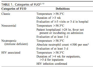
On Friday we spoke about a case of fever of unknown origin or FUO.
The original definition of FUO as proposed by Petersdorf and Beeson in 1961 required the following criteria:
(1) Fever of >38.3 on more than one occasion
(2) Fever for more than or equal to 3 weeks
(3) No etiology discovered after at least 1 week of inpatient investigations

However, the definition has evolved with the increasing population of patients with HIV and neutropenia and the ability to perform investigations in an outpatient setting. The following is a new definition proposed in 1991 by Durack and Street:

Hayakawa et al. Am J Med Sci. April 3. 2012 Epub ahead of date

A thorough history, including onset, duration and nature of the fever, is crucial. While the pattern of fever has not been found to correlate significantly to the etiology, two patterns have been described: (1)Pel-Ebstein (fever for 3-10 days on, then 3-10 days off, typical of Hodgkin's lymphoma) (2) Typhus inversus: reversal of normal diurnal pattern seen in TB.

In many cases the etiology can be narrowed down based on the patient's social history. Including the patient's country of origin, vaccination status, recent travel history (where exactly), animal or insect exposure, recreational activities (gardening or swimming in fresh water), sexual activity, use of recreational drugs. The most at risk population for acquiring an travel related infectious disease, are those who immigrated long ago and return to their country of origin to "visit friends and relatives" (VFR), as these people tend to be the population who take the least precautions.

The following is a table of common causes of FUO:


Hayakawa et al. Am J Med Sci. April 3. 2012 Epub ahead of date

For a comprehensive approach to FUO refer to the following interesting articles:
http://archinte.jamanetwork.com/article.aspx?articleid=215227

Thursday, July 5, 2012

This morning we discussed a case of a 60 year old male with end stage liver disease secondary to Hepatitis C who presented with confusion. Dr Panisko guided us through the process of building a differential diagnosis using the patient's past medical history as a starting point. This is a useful method of organizing your approach, especially when faced with a non-specific presentation such as confusion. Another useful acronym for an approach to delirium is DIMS:

D - Drugs, drugs, drugs
I - Infection
M - Metabolic derangement (endocrine, electrolyte)
S - Structural (intra-cranial process, liver/renal/heart failure)

 Inouye SK. Delirium in older persons. N Engl J Med. 2006;354: 1157-1165

Our patient had multiple possible causes for his confusion:

1) Drugs: Drugs are the most common causes of delirium. Common culprits are psychotropic meds (anxiolytics, antipsychotics, dopamine agonists), anticholinergics, analgesics (opioids), corticosteroids, anticonvulsants etc. Our patient was on seroquel, paroxetine and bupropion, all of which may have contributed to his delirium. One must think of serotonin syndrom given this patient's use of an SSRI. The patient however did not fulfill the Hunter criteria for serotonin syndrome, which includes history of ingesting a seritonergic agent and one of:
  • Spontaneous clonus
  • Inducible clonus PLUS agitation or diaphoresis
  • Ocular clonus PLUS agitation or diaphoresis
  • Tremor PLUS hyperreflexia
  • Hypertonia PLUS temperature above 38ÂșC PLUS ocular clonus or inducible clonus
2) Hepatic encephalopathy: Our patient had allegedly stopped his lactulose which puts him at risk for developing hepatic encephalopathy. The pathophysiology of this involves increased blood to brain transport of neurotoxins (such as amonia, amino acids) and biochemical alterations in the uptake and function of neurotransmitters. Up to Date has a good review on the pathophysiology of HE.

3) Infection: Patients with cirrhosis have altered immune defenses and are considered immunocompromised individuals. Changes in gut motility and increased translocation of bacteria can result in increased risk of developing SBP and other infections. Early treatment of infection is important in end stage liver disease (Management of bacterial infections in cirrhosis, J Hepatol. 2012;56 Suppl 1:S1-12)

4) Hyponatremia: Patients with cirrhosis are at risk for developing hyponatremia. This is largely secondary to the systemic vasodilation resulting in the activation of the renin-angiotensin system and ADH, thereby decreaseing the kidney's ability to excrete free water. Treatment for hyponatremia in cirrhosis involves fluid restriction and vasopressin receptor antagonists.

Physical Exam


We reviewed the physical exam for ascites. The following is a table from the JAMA Rational Clinical Exam series for ascites:

JAMA. 1992. 267 (19): 2645-2649
Bottom line:
1. Sensitive tests for Ruling OUT ascites:
  • Negative history for ankle swelling/increased abdo girth
  • Lack of bulging flanks, flank dullness or shifting dullness
2. Specific tests for Ruling IN ascites:
  • Positive shifting dullness and fluid wave