Hi all. This Dr. C filling in for Natalie today. We had great discussions at Rapid Fire Morning Report today.
On the topic of heuristic thinking and biases here is the reference to the book Dr. Abrams brought up by Daniel Kahneman. It's a fascinating read.
We also discussed bronchiectasis and the choice of antibiotic treatment in an exacerbattion. As Dr Sibbald mentioned, the British Society Guidelines recommend Amoxicillin as first line (often in high doses), with the caveat that previous cultures should guide therapy. This is especially true for coverage of Pseudomonas. We did not delve into treatment of non tuberculous mycobacteria, but here is a BTS reference as well as a more recent ATS guideline for further reading.
Lastly we had a great discussion on managing hyperkalemia. Here is a link to an interesting article that highlights an important point that came up in our discussion:
- Up to 50% of patients with K>6.5 may have no ECG changes!! Often this is related to chronicity, with slower rises in potassium causing less ECG changes, however that level of hyperkalemia requires prompt treatment.
Finally take a look at the CMAJ article on treatment of hyperkalemia from 2010.
Cheers
Thursday, March 28, 2013
Wednesday, March 13, 2013
Painless Jaundice!

This morning we discussed an interesting case of a patient with painless jaundice after receiving multiple courses of antibiotics, including septra, for a presumed cellulitis.
Here is an approach to painless jaundice (not an exhaustive list):
1) Prehepatic causes:
- Hemolysis: i.e. AIHA, G6PD with offending drug
- Impaired hepatic bilirubin conjugation: Gilbert's, Crigler Najjar
2) Intra-hepatic causes:
- Inflammatory: Primary biliary cirrhosis, primary sclerosing cholangitis
- Malignant: Cholangiocarcinoma, hepatocellular carcinoma
- Infectious: Chlonorcus senensis (Chinese liver fluke), Ascaris lumbricoides, Viral: HBV, HCV, EBV, CMV (but usually produces more hepatocellular picture rather than a cholestatic picture), alcoholic hepatitis
- Drug induced liver injury
- Other: NASH, liver infiltration (amyloid, sarcoid, lymphoma)
3) Post-hepatic causes:
- Cholelithiasis: common bile duct obstruction, Mirizzi syndrome where distended gallbladder causes obstruction of extrahepatic bile duct
- Pancreatic neoplasm
- Stricture post instrumentation
A little more on Drug Induced Liver Injury (DILI)
1) Types of DILI:
- Can mimic all patterns observed in primary liver disease
- Acute hepatocellular injury: increased ALT/ALP ratio
- Cholestatic injury: Isolated increase in ALP, with decrease in ALT/ALP ratio
- Granulomatous hepatitis
- Steatohepatitis
- Can present as acute or chronic liver injury
- Acute hepatocellulary injury and fulminant liver failure:
- Examples of drugs that cause acute fulminant hepatitis include: acetaminophen, isoniazid, sulfonamides, cotrimoxazole, ketoconazole, anticonvulsants
- Acute Cholestatic injury:
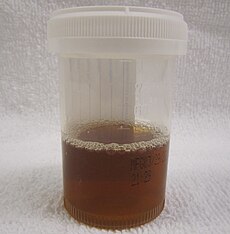
- Presents clinically with jaundice, pruritis, dark urine and pale stools
- Liver enzymes reveal elevated ALP, GGT and Bili.
- Common culprit drugs include: anabolic steroids, oral contraceptives, prochlorperazine, and antibiotics (septra and erythromycin)
- See the following articles for more information:
- NEJM article on DILI
- Case report of Septra induced prolonged cholestasis: http://www.ncbi.nlm.nih.gov/pubmed/1587437
Thursday, March 7, 2013
Use of Fluoroquinolones

Today in morning report we had a quick discussion on the use of fluoroquinolones. Here are some pearls to consider prior to prescribing a fluoroquinolone:
- Sensitivity to UV: wear sun protection!
- Prolongation of QT interval: Check baseline ECG and repeat ECG while on medication if baseline QTc is prolonged
- Tendonopathy and tendon rupture: worse in elderly >60yrs, mainly Achilles tendon
- Rash: Immediate IgE mediated reactions and delayed type hypersensitivity reactions (maculopapular rash)
- Hepatic toxicity: may cause transaminitis and rarely acute liver injury
- Hypoglycemia: mainly seen with gatifloxacin
- Exacerbation of myasthenia gravis
Wednesday, March 6, 2013
Surviving Sepsis Campaign

The Surviving Sepsis Campaign came out with updated guidelines for the management of severe sepsis and Septic shock in 2012. Here is a summary. Please refer to this link to access the entire article: Surviving Sepsis Campaign
A. Initial Resuscitation:
- Recognize septic shock: defined as documented hypotension despite adequate fluid challenge and evidence of end organ hypoperfusion (lactate >4g, AKI, change in LOC, cardiac ischemia)
- Goals for the first 6 hrs:
- CVP 8-12mmHg
- MAP 65mmHg or greater
- Urine output goal 0.5mL/kg/hr
- Central venous saturation 70% or mixed venous O2 sat of 65%
- Target resuscitation to the normalization of lactate
B. Diagnosis:
- Septic w/u: cultures should be drawn prior to antibiotic administration anaerobic/aerobic at least one peripherally and one from all central lines
- If fungal infection/candidiases is suspected: use 1,3 beta-D-glucan assay, mannan and anti-mannan antibody assays
- Imagine as needed to investigate source
C. Empiric Therapy
- Administer IV abx within first hour of recogntition of shock.
- Think about potential bugs and penetration of IV abx
- Combination therapy for severe sepsis/neutropenic patients is recommended for up to 3-5 days but should be narrowed thereafter. Consider combo therapy for pseudomonas, acinetobacter and other multi-drug resistant organisms (i.e. an extended spectrum beta lactamase and gentamycin or flouroquinolone)
D. Get Source Control
- Every effort should be made to get source control within 12 hrs of identification, this may involve getting the surgeons or interventional radiology involved. Abx treatment should be limitted to 7-10 days, unless there was slow response to abx
- For example, infected peripancreatic abscess or infected necrotizing pancreatitis should be drained percutaneously or surgically once viable and non-viable tissue is demarcated.
- If percutaneous devices are a possible source infection, they should be removed promptly!
E. Infection Prevention:
- Selective oral and or digestive decontamination should be used i.e. oral chlorhexidine
Wolfram's Syndrome and secondary causes of diabetes

Today we had an interesting discussion of a patient with Wolfram's Syndrome or DIDMOAD, this generated an interesting discussion on secondary causes of insulin resistance:
1) What the heck is DIDMOAD?
- Wolfram syndrome has three genetic forms one of them being DIDMOAD
- It is an autosomal recessive disorder with incomplete penetrance, affecting 1/770 000
- DI - Diabetes insipidis: due to loss of vasopressin secreting cells, anterior pituitary dysfunction has also been described
- DM - Diabetes Mellitus: Patient typically develop diabetes in childhood requiring insulin (due to an inability to convert pro-insulin to insulin)
- OA - Optic atrophy occurs early in childhood
- D - Deafness: sensorineural deafness
2) What are other causes of insulin resistance?
- Primary pancreatic: Infiltrative (hemochromatosis, amyloidosis), cystic fibrosis, pancreatic cancer, chronic pancreatitis, surgical removal/trauma
- Endocrine: Cushings (secondary to elevated cortisol), acromegaly, pheochromocytoma (secondary to adrenergic stimulation), Conn's (secondary to hypokalemia)
- Drugs: Steroids (increases gluconeogenesis and decreases peripheral glucose transporter), thiazide diuretics, atypical anti-psychotics, nicotinic acid, HAART therapy, ocreotide infusion, inotropes
- Unusual causes: muscular dystrophy, friedrich's ataxia, Wolfram's syndrome etc.
3) Work up and Management of hyperglycemia:
- Confirm: Repeat the accucheck
- Look for causes:
- Known diabetes? HHS/DKA (look for precipitating factors: infarction, infection, insulin non-compliance, new diagnosis of diabetes)
- Secondary causes of insulin resistance: i.e. medications
- Look for perpetuating factors:
- Severe hypovolemia results in both hemoconcentration and decreased GFR leading to decreased filtration of glucose
- Increased adrenergic/stress state drives gluconeogensis and insulin resistence
- Investigations:
- Physical exam to look for precipitating factors and degree of volume depletion
- Laboratory work to look for DKA/HHS and precipitating factors:
- ABG, anion gap, serum ketones, serum osmolarity, electrolytes (esp. K+), creatinine
- Septic w/u, ecg, troponin/ck, amylase/lipase
- Treatment:
- Fluids for the hyperglycemia
- IV Insulin to close the anion gap and decrease fat metabolism and the generation of ketones. Continue until the anion gap is closed
- potassium replacement
- Treat the underlying cause
- Transition to SC insulin once patient's B.G. is stable and insulin requirements have been minimal and stable and patient is eating... and in the daytime! Ensure at least 1-2 hrs of overlap.
- Please see the following article for treatment of Diabetic ketoacidosis: CMAJ 2003 Treatment of DKA/HHS
Subscribe to:
Posts
(
Atom
)
