Thursday, August 15, 2013

Scleroderma

Derived from the term "scleros", meaning thickened, scleroderma is a heterogeneous disease that can effect the skin and multiple internal organs. The terminology is usually categorized as systemic versus localized sclerosis, and within systemic sclerosis there is limited and diffuse disease, which is based on cutaneous distribution.

Localized scleroderma can be subdivided in linear and morphea:

Linear scleroderma - "coup de sabre", which gets its name because it appears like a scar from a blade. This is usually limited to dermatomal distribution and is common in younger patients.

Morphea - can be circumscribed or generalized or focal. There is significant variability within this group, with multiple subtypes. Morphea often spares the face, which is helpful in separating this from systemic sclerosis.

Systemic scleroderma

Limited cutaneous systemic sclerosis (lcSSc) - have cutaneous changes limited to the hands and forearms, on formthat is well described is CREST:
1. Calconisis cutis
2. Reynauds 
3. Esophageal dysmotility
4. Sclerodactyly
5. Telengactasia

Diffuse cutaenous systemic sclerosis (dcSSc) can have additional clinical manifestations including:

1. Vascular abnormalities: Reynauds
2. MSK: myalgias, arthralgias, fatigue. Inflammatory arthritis is uncommon. Osteolysis and bone destruction can occur.
3. Pulmonary: interstitial lung disease or pulmonary hypertension can develop. This can be quite debiliatating for patients. 
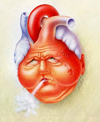
4. Cardiac: arrhythmias, pericardial and myocardial disease. Myocardial disease is felt to be secondary to small vessel spasm, similar to Reyanaud's phenomenon. 
5. Renal: "Renal crisis", with azotemia, hypertension and bland urine sediment. 
6. Neurologic: multiple different types of neuropathies have been described, with autonomic, peripheral, cranial and entrapment documented.

Another entity is systemic sclerosis sine scleroderma, which has internal organ manifestations without skin findings.

As mentioned today, there is an increased risk of malignancy, both lung and GI, though the mechanism for this is unclear. Smoking was found to be a risk factor in previous studies.

The case discussed today involved a patient with systemic sclerosis with acute on chronic kidney injury, which always raises the possibility of renal crisis. This is defined by an acute, progressive worsening of renal failure, increased blood pressure and bland urine sediment (mild proteniuria with few casts/cells). Up to 80% of patients with SSc will have renal involvement, with only 20% experiencing renal crisis. Average BP in these patients was found to be ~ 175/105, with signs suggestive of hypertensive emergency (encephalopathy). Additional tesing can reveal microangiopathic hemolytic anemia, with schistocytes and low platelets. Risk factors include severe skin disease, corticosteroid use, cyclosporine use, and anti-RNA polymerase antibodies. 

Wednesday, August 14, 2013

IgG4 disease

An common dilemma in medicine is distinguishing between worsening autoimmune disease and infection in patients with chronic autoimmune disease when they present with worsening symptoms. Increasing dyspnea may represent a worsening inflammation in lungs, versus a pulmonary infection secondary to immunosuppressant drugs. In a recently discussed case,  we encountered this scenario in a patient with IgG4 disease (pancreatitis and sclerosing cholangitis) and a positive cytomegalovirus PCR in bronchoalveolar fluid lavage.

IgG4 related disease is relatively new entity, first being recognized in 2003. Initially felt to be solely with autoimmune pancreatitis, it has now been identified to cause disease in nearly all organs. Its involvement in disease development is currently unclear, where its role as a primary or secondary factor in pathogenesis is yet to be confirmed. Interestingly IgG4 levels in serum and tissue are helpful but not necessary (negative ~30%) to confirm a diagnosis, where additional pathologic features can be helpful. Clinically, patients tend to present with subacute disease involving one ore more organs. Fever and elevated inflammatory markers are uncommon. Organ sclerofibrosis with tumour like lesions and atopic symptoms (asthma, allergy) are a common manifestations. A list of organs that can be involved are listed from a recent NEJM review:

1. Mikulicz’s syndrome (affecting the salivary and lacrimal glands)
2. Küttner’s tumor (affecting the submandibular glands)
3. Riedel’s thyroiditis
4. Eosinophilic angiocentric fibrosis (affecting the orbits and upper respiratory 7.tract)
5. Multifocal fibrosclerosis (orbits, thyroid gland, retroperitoneum, mediastinum etc)
6.Inflammatory pseudotumor (affecting the orbits, lungs, kidneys, and other organs)
7. Mediastinal fibrosis
8. Retroperitoneal fibrosis (Ormond’s disease)
9. Periaortitis and periarteritis
10. Inflammatory aortic aneurysm
11. Tubulointerstitial nephritis with extensive tubulointerstitial deposits

Treatment can be monitoring (in slowly progressing asymtpomatic) or involve immunosuppressants. Prednisone is the mainstay with slow tapers over months +/- sparing agents like azathiaprine, methotrexate or mycophenylate. Rituximab use has also been reported. 

Given the patients medications, infection was considered as the underlying cause, specifically CMV pneumonitis. CMV infection most commonly occurs in the GI tract and lungs, when clinically significant. There are multiple ways to test for infection, including serology, cultures, antigen detection and molecular amplification. There are no consensus guidelines for the diagnosis CMV pneumonitis. PCR testing is quite sensitive, but there are no cut-off levels for diagnosing pneumonitis, where low levels are not interpretable. High levels in the appropriate clincal context chould prompt consideration of gancylovir treatment. CMV is often detected in patients with autoimmune disease without clinical symptoms on BAL (~20% -CHEST 2001). In patients with pneumonitis (dyspnea, hypoxia, infiltrates) immunostaining for CMV in BAL samples has a high sensitivity and specificity and is useful test in confirming disease. This is a challenging scenario and may require further discussion with both ID and respiratory medicine specialists. See below for additional information. 


Friday, August 9, 2013

COPD exacerbations and Antibiotics

In the current era of antimicrobial stewardship, select use of antibiotics is increasingly important. Physicians are aware of the numerous side effects of antimicrobial therapy, increasing resistance patterns and additional costs associated with prescribing, and hence try to target only those patients who will benefit from these medications because of an underlying infection. Infections are felt to cause up to 75% of all exacerbations, and can be due to bacteria, virus' or additional infectious agents. In 2002, a study in NEJM showed that exacerbations were not only associated with bacterial infections, but with new organisms as opposed to those previously seen in colonization.


Antibiotic therapy in COPD exacerbations has been a controversial topic. Ignoring the early, statistically flawed, small sampled trials from the mid-20th century, Anthonisen et al. performed a RCT in 1987 looking at antibiotics in COPD exacerbation. They categorized patients based on the presence of sputum purulence, increased sputum volume, and increased dyspnea. Patients with all of these characteristics had the largest benefit with antibiotics compared to placebo with an >15% reduction in deteriorations. As the number of criteria reduced, there was less of an effect between the antibiotics (doxy, septra or amox) vs placebo. This is why these three qualifiers have become engrained into our COPDE history and used in the consideration of antimicrobials. The GOLD guidelines recommend antibiotics for exacerbations with sputum purulence with increased volume or dyspnea. The severity of illness also plays a role in considering antimicrobial therapy. A Cochrane Review found that patients admitted to the ICU have a significant benefit from antibiotics therapy, but for inpatients without ICU admission the benefits was not as clear. A retrospective study published in JAMA (2010), examined over 80,000 patients who were hospitalized for COPDE. Failure to receive antibiotics in the first 48 hours was associated with increased mortality, repeated admission and mechanical ventilation.

Use of antibiotics for outpatients has less clinical evidence. There have been randomized trials showing showing decreased duration of symptoms (by days) and longer times until repeat exacerbation with the use of Amox-Clav. However, given the individual risks of harm and mild benefits with antibiotics, utility is questionable. Amox-Clav has been compared to other antibiotics (moxi), without showing any significant difference. Duration of five days may be an appropriate length of treatment in the outpatient setting, where a meta-analysis in COPD showed that five days compared to seven days showed no differences in treatment success and lower adverse events.

Overall, there is no specific antibiotic regiment that should be chosen, and patients should have antimicrobials based on their previous history, local resistance patterns and risk factors for pseudomonas. Whether or not antibiotics should be given at all should be based on an individual basis. I also consider additional clinical factors used in the diagnosis of community acquired pneumonia, such as presence of fever and focal consolidation on chest x-ray.

Anthonisen Annals

Thursday, August 8, 2013

Candidemia

A recently discussed case was of a patient with sepsis secondary to fungemia with Candida parapsilosis. Candida in the blood should never be assumed to be contaminant, though this can sometimes be a result of a contaminated line. As with all cases of sepsis, Candidemia has been increasing in the US and Canada.

Candidemia can be invasive or non-invasive, the presence of end organ involvement (endophthalmitis, endocarditis, renal and CNS involvement) defines invasive disease. Diagnosis should be based on positivie blood cultures. Skin biopsies if lesions are present can also be used to identify systemic disease. Recently, assay for beta-D-glucan, a component of the cell wall of Candida has been shown to be useful in diagnosis candidemia. This test is not specific to Candida and will be positive in other invasive fungal infections. This test has been shown to have reasonable sensitivity and specificity for fungal infections. Risk factors identified by several case-control studies in hospitalized and ICU patients included the following:


1. Hickman lines
2. ESRD on dialysis
3. Broad spectrum antibiotics use
4. Gastric acid suppression
5. ICU admission
6. Total parenteral nutrition (present in our patient)
7. Immunsuppression


Candida albicans is the most common species identified in candidemia, however additional strains are being recognized with increasing frequency. C. glabrata, C. parapsilosis, C. krusei and C. tropicalis are amongst the most common species seen. C. glabrata can be resistant to azole therapy, while all C. krusei is resistant to this class of medications. A less common species C. lusitaniae can develop resistance to amphotericin, which should be recocgnized by treating physicians.

Empiric treatment of fungemia should consider multiple factors: history of recent azole antifungals, nuetropenia, severity of illness, local epidemiology, invasive disease and history of resistant organisims in the individual. If the patient was well and had few risk factors, initial treatment with fluconazole IV is reasonable. otherwise, neutropenic/septic patients should receive an echinocandin (caspofungin/micafungin) until speciation occurs. Amphotericin has significant toxicity and tends to be used less. Duration of therapy is based on consensus guidelines, and the IDSA has said 2 weeks following negative blood cultures is a reasonable duration in patient without invasive disease (abscess/endocarditis).

See the link below for candidal guidelines:

IDSA guidlines for candidemia


Wednesday, August 7, 2013

Myasthenic Crisis

Myasthenic crisis is a worsening of muscle weakness precipitating respiratory failure and need for ICU admission and intubation in patients with myasthenia gravis (MG). MG is an autoimmune disorder characterized by antibodies directed against acetylcholine receptors. This results in diffuse muscle weakness, primarily involving bulbar, peripheral and respiratory muscles. Although MG is a rare condition (~5-10/100,000), approximately 15% of patients will develop a crisis during the course of their disease. Despite advances in understanding disease pathogenesis and treatment options, myasthenic crisis carries a mortality of up to 8%. Patients clinical condition can worsen precipitously, sometimes as a result of a triggers and other times without clear cause. Precipitants include:


1. Infection -  usually pnuemonia/aspiration (~70% of causes for crisis)
2. Medications - Tapering of steroids, acetycholinesterase. Or worsening muscle dysfunction from additional medications: anaesthetics, antibiotics, anticonvulsants, antiarrhythmics, beta blockers, magnesium and many others
3. Surgery
4. Pregnancy and childbirth

The cholinergic crisis (worsening of muscle weakness from toxicity due to acetylcholinesterase inhibitors (AChEI)), should be considered in pateints as it can look similar and has a very different treatment approach. Patients on less than 120mg of pyridostigmine q3h are reported to be at low risk of developing this problem.

Investigations that can aid in the diagnosis of myasthenic crisis include: ABG, CXR, vital capacity (VC) and mean inspiratory pressure (MIP). As weakness worsens, an ABG will reveal hypoxemia and hypercarbia. Imaging can identify precipitating infections and alternative causes of dyspnea. A VC of less than 1 litre or 20cc/kg is concerning of imminent failure and warrants intubation. MIP's of less than -30cmH20 should raise concern and lead to endotracheal intubation.

There are several treatment options for myasthenic crisis. These include:

1. AChEI's
2. Steroids
3. Plasmapheresis
4. IVIg

The use of AChEI's in a crisis state is controversial. The awareness of cholinesterase crisis has led many physicians to monitor patients for several days before increasing these drugs. Many prefer to hold these medications and monitor the patient clinically to try and identify the underlying cause, while others continue the medications. There have been randomized studies comparing AChEI +/- steroids, or plasmapheresis, which did not show any additional benefit in patient outcomes.

There is not a large amount of evidence for increasing steroid dosage in acute crisis. Guidelines suggest 60-80mg of prednisone a day and monitoring for acute myopathy from this medication. This should be considered primarily in patients with severe disease in the ICU setting.

Plasmapheresis and IVIg have been promoted as both first line therapies in myasthenic crisis. This idea is that removal or disruption of autoantibodies would result in improvment of muscle function. Both of these treatment options have realatively quick onset of action (days), but do not result in persistent improvement (weeks). Head to head studies have suggested that plasmapheresis has better short term response, but increased side-effects compared to IVIg. Long-term therapy should be considered in conjunction with acute management given this can take time to work (i.e. azathioprine), and the acute therapies will have worn off.

See link below for more info:

Myasthenic crisis


Thursday, August 1, 2013

Pel-Ebstein Fever

This eponym has been raised several times in the month of July in regards to patients presenting with either fever of unknown origin or fever in the context of a previous malignancy. Pel-Ebstein fever refers to a cyclical fever in the context of Hodgkins lymphoma (HL). The description is of fevers lasting days to weeks with defervescence of similar duration. The duration of the absence of fever may decrease as disease progresses. Pal and Ebstein described this separately in two publication in the mid 1880's. Editorials have been published commenting on the original articles, which recognize that the prevalence of HL was not confirmed in many of the subjects described in the original article. It has also become more recognized that other causes of fever can produce a similar pattern, where case reports of CMV and tuberculosis report cyclical fevers (Schattner 2010.). The diagram posted is from a NEJM clinical image from 1995 which charts the fevers of a patient with HL, demonstrating the classic pattern. How often patient with HL present with this pattern is unclear, but its thought to be rare. If nothing else, the eponym has labelled this phenomenon allowing it to be remembered, which may be the only reason its discussed. There are other systemic manifestations of lymphoma that are just as interesting, but don't seem to have the same notoriety.

Alcohol induced pain, usually at the site of involved lymph nodes or bony disease was first described in the mid 20th century. Its reported to occur quickly after the ingestion of alcohol in  less than 10% of patients. Pruritis is a common manifestation of lymphoma, where ~10-15% will experience this, sometimes being quite severe. It may be an early presenting symptom and when severe may carry some prognostic value. I would argue that pruritis is less commonly mentioned as a symptom of HL than pruritis, and may be a more valuable piece of knowledge.

Wednesday, July 31, 2013

Non-tuberculous mycobacterium


With the development of improved microbiologic techniques, there has been increased recognition and identification of non-tuberculous mycobacterium (NTM). Over the past several decades we have accumulated over 100 species of this bacterial group. These organisms are identified through positive acid-fast staining, culture and speciated with 16S ribosomal rRNA sequencing. These bacteria are ubiquitous in the environment living in the soil and water sources. Interestingly, there does not seem to be person to person transmission, which is a major risk factor for other bacterial transfer in certain populations (i.e. cystic fibrosis and B.cepacia). Mycobacterium can be categorized as following:


1. Mycobacterium tuberculosis - TB
2. Mycobacterium leprae - Leprosy
3. Slow growing non-tuberculous mycobacterium - (M. avium, M.kansasaii, M. xenopi, etc.)
4. Rapid growing non-tuberculous mycobacterium - (M. abscessus, M.fortuitum, etc.)


Of the NTM, Mycobacterium avium and kansasaii are the most prevalent, though M. xenopi is common in the province of Ontario (~18% of NTM).

Clinical manifestations of NTM fall into several catgeories:

1. Pulmonary disease - most common manifestation of NTM, often associated with COPD, bronchiectasis, old TB with structural lung disease and cavities. Most common in males, age >50. Presents as chronic cough, sputum production, constitutional symptoms. Imaging studies can reveal consolidation and nodules. Again, these infections are often associated with structural lung disease/cavitation. Subacute pulmonary hypersensitivity can be seen in exposure to M. avium, referred to as  "hot tub lung". This is when the exposure is from a large inoculum of organism present uncleaned water from hot tub. It presents with subacute dyspnea, fever, cough, infiltrates and sometimes respiratory failure.

2. Disseminated NTM - seen in immunosuppressed patients, usually HIV. Most common organsims are M. avium and M. kansasaii. Presentation is dominated by fever and other constitutional symptoms. Organomegaly and abdominal tenderness may be seen.

3. Skin/soft tissue/bone disease - associated with catheters and following procedures with break the skin. The organisms involved are M. abscessus, M. chelonae, M. fortuitum and M. marinum. These require source control and antibiotics.

4. Lymphadenitis - more common in children. Present with lymphadenopathy with purulent drainage. M. avium is the most common organism

The case we discussed today was of M. avium in a patient with COPD and cavitary disease. They were taking a macrolide, fluoroquinolone, rifampin and ethambutol. Pateints with bronchiectatic disease without cavities may manage with ethambutol and macrolide alone. The multiple drugs used in our patient was likely related to cavitary disease. The IDSA has guidelines on the management of NTM, which can help guide therapy. Its a challenging disease to treat, which usually requires at least one-year of antimibrobial therapy following negative cultures (hence it often is longer). Monotherapy with macrolides should be avoided to prevent resistance development. Intravenous regiment with amikacin or carbapenems are sometimes required and carry signficant toxicities. Different species  require different regiments and resistance patterns should be considered. Patients can from surgery if there is local disease (specifically with M. abscessus).

See the following link for the guidelines
NTM IDSA guidelines

Thursday, July 25, 2013

Brain Natriuretic Peptide

Brain natriuretic peptide (BNP) is named as such, because it was initially identified in brain tissue, however it's primarily released by the myocardium. There are two peptides produced during processing, the amino-terminal BNP and the biologically active BNP. Along with atrial natriuretic peptide (ANP), BNP is involved in salt and water balance in response to myocardial stretch from increased filling pressures. It's influences lead to diuresis, natriuresis and hypotension.

Clinical evaluation of these hormones can be performed using multiple assays. There are tests available for both NT-BNP and BNP, some of which are rapid point of care testing, which can be used in the emergency department. It should be recognized that these assays have different cut-offs for normal values and can be influenced by age, gender and renal failure (higher in elderly and women, low in obesity). There have also been studies in specific ethnic groups which have identified the different reference ranges for certain ethnicities (Choi, 2007). Chronic congestive heart failure may produce persistently elevated levels, making it difficult to interpret spot samples without documented baselines.

In 2002, the Breathing Not Properly study (BNP), evaluated patients presenting to hospital with dyspnea, documenting BNP levels on all patients and comparing level in those eventually felt to have CHF versus alternative diagnoses. Patients with CHF had levels over 400pg/ml with high specificity,  while levels less than 50pg/ml  had a negative predictive value of 96%. Higher levels were also found in patients with atrial fibrillation and renal failure. The authors felt BNP was superior to any one individual clinical factor in predicting CHF exacerbation. A similar trial (PRIDE), examined the ability for NT-BNP to predict CHF in acute dyspnea. They found that levels over 450pg/ml (in patients under 50) and over 950pg/ml (in patients over 50) were sensitive and specific for CHF. NT-BNP was superior to clinical judgement in predicting CHF in this trial. The use of BNP markers has been shown to decrease ICU and hospital duration of stay by ~ 24 hours but have no statistically significant impact on overall mortality (Porapakkham, 2010). 

Despite this information, laboratory testing shouldn't replace clinical judgement, as individual variability contributes to lab interpretation. BNP is however a useful marker in the setting of evaluating acute dyspnea, and there is some additional literature suggesting it may be useful in the outpatient setting, where levels may predict mortality in chronic CHF and be used as a target for treatment.

Cardiac biomarkers NEJM

Heroin

Like many of the drugs used today, heroin is a chemically manufactured and not naturally occurring. Given its relation to organic molecules derived from opium (ie. morphine/codeine), it belongs to the opioide family. This differs from opiates, which are naturally occuring molecules originally derived from the poppy plant. With a simple acetylation reaction (the same synthetic step used to create aspirin from its parent molecule), heroin was born from morphine, and the rest is history. Shortly after its creation it was marketed over the counter as an analgesic. Acetylation is maintained when drug is injected (bipassing the first pass effect) and is what allows fat solubility and movement into the central nervous system.

Heroin use as a recreational drug has been a major health concern for nearly a century, and despite widespread knowledge of its effects, use continues to grow. Sequlae of heroin use includes, overdose, poisening from contaminants, endocarditis, injection site abscesses, renal disease and dependence amongst other things. A publication in The Lancet (2007) identified heroin as having the highest risk of harm to users compared to 20 other illicit substances. The concern of health consequences is what has driven many cities to initate clean needle injection sites. A recently discussed case highlighted the importance of considering the presence of adulterants in heroin, which can have significant consequences.

A review published in 2011 examined the content of heroine samples acquired over a 5 year period. Heroin was found to make up only ~25% of heroin samples, where additional substances made up the rest of the product. Tylenol and caffeine were the most common additives, though levamisole (an anti-helminthic medication) has been identified previously. Levamisole was report to be present in up to 4% of cocaine samples in a European study (Schneider et al.), though much higher values have been identified in US publications (upwards of 70% of samples, JAMA). Ingestion can result in profound neutropenia, leukoencephalitis and vasculitis. Physicians should recognize the heterogeneity of illicit substances and look for symtpoms which may be consistent with their toxicities when patient present with intoxication.

Heroin in Toronto

Thursday, July 18, 2013

Steroids for Alcoholic Hepatitis

In 1978, Maddrey and colleagues published a randomized trial of prednisolone versus placebo in alcoholic hepatitis. They found the laboratory markers (bilirubin/prothrombin time) could predict which patients would benefit from corticosteroid therapy, where patients with a score >32 had a mortality benefit at thirty days. Thirty-five years, and over a dozen RCT's and meta-analyses later, the use of steroids remains controversial.


Prednisolone is the most widely studied corticosteroid and is preferred over prednisone, considering prednisone is converted to prednisolone following hepatic metabolism. Forty milligrams per day is the most widely used dose and is continued for 28 days duration, followed by a 2 week taper. The populations studied tended to have isolated alcoholic hepatitis, and patients with GI bleeding, pancreatitis and active infection tended to be excluded from study. The small sample sizes and heterogeneity of studies has made it difficult to determine the effect of pharmacotherapy. A systematic review found no mortality benefit in patients with mild to moderate hepatitis, but found a mortality benefit with steroids in those at highest risk (Rambaldi et al. 2008). Interestingly this therapeutic effect may have a threshold, where patients with a Maddrey descrimant function score (MDF) >54, were found to have increased mortality with steroids.

Given a mortality rate of over 30% in patients with severe alcoholic hepatitis, combination therapy trials have been designed with the hope of improving outcomes. Prednisolone +/- N-acetylcysteine (NAC) were compared in a randomized control trial published in NEJM (Nguyen-Kach et al. 2011). There was no significant difference in mortality at 6 months, but there was improvement in survival at 1 month. Patients in the combination group were also less likely to develop hepatorenal syndrome and infection. Recommendations for NAC were not included in the recent update of the American Association for the Study of Liver Disease (AASLD) guidelines.

An additional score (Lille score) was developed and found to be helpful in determining if patients with alcoholic hepatitis were to be responsive to corticosteroid therapy. This score predicted six month mortality by comparing using laboratory data over time. Patients with chronic liver disease often have multiple causes contributing to disease progression. It is important to take a detailed alcohol history and consider pharmacotherapy in the appropriate clinical context.

Steroids and NAC - NEJM


Wednesday, July 17, 2013

Staphylococcus aureus bacteremia

Methicillin sensitive staphylococcus aureus bacteremia (MSSA) is a life threatening condition. Even in the era of antimicrobial therapy, the overall mortality of approximately 30%. Once recognized, clinicians should focus on risk stratification and prevention of systemic complications. This is the reason for automatic infectious disease consultation following a positive blood culture with S. aureus.

Prior to speciation, gram stain results will become available and show gram positive cocci in clusters. This warrants the initial treatment with vancomycin, given methicillin resistant S. aureus (MRSA) is a possibility, which would not respond to traditional beta-lactam antibiotics. Risk factors for MRSA amongst patients with S. aureus bacteremia include previous colonization, nosocomial infection, older age, admission from long term care facility and history of broad spectrum antibiotics. Intravenous drug use has actually been found to be less associated with MRSA, though there have been conflicting results (JAMA Roghmann). 

Once identified as MSSA, patients should be switched from glycopeptide antibiotic (vancomycin) to beta-lactam (cloxacillin/ancef), given an identified mortality benefit in previous trials. The risk of complicated infection (systemic/metastatic infection - endocarditis, discitis, meningitis etc.), can be predicted using clinical and laboratory data. Fowler et al. found that community associated infections, fever at 72h post antibiotics, persistent blood culture positivity at 72h post antibiotics and skin changes suggestive of acute infections predict complicated bacteremia. This may lead to alterations in duration of antibiotic therapy, which is typically a minimum of two weeks. Predicting infectious endocarditis in these patients and need for echocardiography has been associated again with persistent bacteremia and presence of intracardiac devices.

See the link below to Fowler's article.


Friday, July 12, 2013

Myositis and malignancy

In 1975, Bohan and Peter proposed a diagnostic classification system for polymyositis (PM) and dermatomyositis (DM), a rare group of idiopathic inflammatory myopathies. They felt the presence of proximal muscle weakness, typical dermatologic manifestations, laboratory markers of muscle damage (high CK), EMG data and features on muscle biopsy would confirm a diagnosis. Today, similar clinical data is used for the diagnosis with the addition of auto-antibodies testing, however these diseases are recognized as having an increased amount of heterogeneity. In 2004, a list of categories including possible and definite DM/PM was created, also recognizing inculsion body myositis and dermatomyositis with a lack of muscle involvement. A simplified breakdown is considering if the patients presentation is highly characteristic presentation, non-specific but typical, atypical or those with skin manifestations without myositis.

An association between malignancy and myositis has been recognised since the early 20th century. It wasnt until large retrospective cohort analyses were performed that this association was confirmed. Approximately 25% of patients with DM have been found to have underlying malignancies, most of which are identified within two years of presentation. PM has a lower risk than DM, but increased from the general population.There are no guidelines on how the screening process for malignancy should take place, just that a focused assessment be performed and age appropriate cancer screening initiated. Additional risk factors for malignancy in myositis include increased age, males and severe cutaneous manifestations. The most common malignancy associated with myositis are adenocarcinomas (~70%), though there are race specific differences. Patients of south-east Asian descent with myositis were most likely to have nasopharyngeal carcinoma as the underlying cancer (~20%). Western studies have identified, ovary, breast and lung amongst the most common malignancies. Features consistent with the anti-synthetase syndrome, including fever, interstiital lung disease, Reynaud's phenomenon, arthritis and anti-Jo positivity are less associated with underlying cancer. The pathogenesis of inflammatory myopathies with underlying cancer is unclear. Case reports have suggested immune mimicry, where cell mediated response against the tumour results in cross reaction with muscle antigens. There have been conflicting results on the response of myopathy following the treatment of the underlying tumour, which may be confounded by the burden of disease at the time of diagnosis.

The main thing is to recall an association between these clinical entities, remembering to perform a focused search for malignancy in patients with inflammatory myopathy.

Thursday, July 11, 2013

Echocardiography and pulmonary embolism


Echocardiography is not recommended routinely as part of the diagnosis of pulmonary embolism (PE). However, given presenting symptoms and clinical instability associated with PE, patients may have this investigation performed during their admission.The availability and accuracy of CT protocols for PE has reduced the need for additional diagnostic testing. As stated, an echo is not the test of choice for PE, considering only up to 40% of patients with PE will have abnormalities. The value of its use may lie within its prognostication.

Signs suggestive of a PE on echo include, RV dilation and hypokinesis, pulmonary hypertension, tricuspid regurgitation and IVC distention. A decrease in LV size can be seen as a results of a dilated RV and bulging of the interventricular septum. Occasionally (~5%), of patients with PE will have an observed thrombus in the RV on echocardiography, confirming the diagnosis. In acute PE, RV dysfunction while sparing the apex (McConnell's sign) may be seen, which has a documented specificity of 94%.

Moderate to severe RV dysfunction on echo has been linked to increased mortality in acute PE. One Swedish study found a 6 fold increase in death compared to patients with normal RV function. The presence of malignancy was also a negative predictive factor in these patients. This finding was identified as one of the most important predictors of increased death at 3 months post presentation. The increase in associated mortality with RV dysfunction suggests this finding may be useful in supporting the decision for thrombolysis in acute PE. The MAPPET registry (see link), found that patients with moderate to severe RV dysfunction have reduced mortality with thrombolysis (with increased risk of severe bleeding), and those with RV dysfunction given anticoagulants alone were at risk for repeat clot. It should be noted, that these patients had normal blood pressures at the time of thrombolysis. RV dysfunction on echo has also been shown to improve post tPA.

The above information makes a case for echocardiography, by a trained specialist, in the patient presenting with symptoms suggestive of PE, adding another diagnostic piece to the initial assessment. It is also valuable in predicting patients with poor outcomes in the months post evaluation.

Acute PE: a multicentre registry

Wednesday, July 10, 2013

Antipsychotics in delirium and dementia


Medication therapy has become commonplace in the treatment of patients with delirium/dementia during hospitalization. Ideally, patient confusion/aggression can be controlled with non-pharmacologic therapy, however when changes in mental status become a threat to patient and health care worker safety, medications can be helpful.

Antipsychotics are generally categorized as first (typical) and second (atypical) generation drugs. As the name suggests, second generation drugs were developed later, where the term "atypical" neuroleptics was introduced in 1998. These were developed to control psychiatric illness, while minimizing side effects. Both classes of drugs antagonize dopamine (D2) receptors, improving hallucinations and delirium. Atypical antipsychotics bind the D2 receptor with less affinity and higher specificity for the mesolimbic area of the brain. This is felt to cause similar efficacy with less extrapyramidal side-effects, which include rigidity, spasticity, and tardive dyskinesia as well as negative symptoms (psychomotor retardation), attributed to blockade in the nigrostriatal and prefrontal regions respectively. 

Neuroleptic medications are amongst the most common drugs used to treat delirium and behavioral dementia. In 2005, the FDA issued a warning statement for these medications as a group, citing a small but significant association with increased mortality with their use. This increased mortality was secondary to an increase in infectious (pneumonia) and cardiovascular (sudden death/CHF/stroke) events. An increase in cerbrovascular events was seen with multiple atypical antipsychotics (ex. risperidone, olanzapine). This has led to controversy regarding the use of these drugs in clinical practice.

Psychotropic medications should be used judiciously in response to delirium, but are sometimes warranted. These drugs should not be provided prophylactically, considering they have not been shown to reduce delirium incidence, but duration following it's occurrence, which was examined in an RCT of postoperative delirium (kalisvaart et al. 2005). The efficacy between agents is similar, where comparisons of haldol (typical) and risperidone, qeutiapine, olanzapine and ziprasidone (atypicals) have been performed. To manage aggressive behavior in patients with dementia, risperidone has been investigated the most and showed mild benefits in multiple trials. The meta-analysis (2005 JAMA), examined 15 RCTs of atypical antipsychotics and placebo in delirium and found a grouped reduction in mortality in the placebo group, however amongst the specific drug subgroups, no increased mortality was identified. Long term follow up patients randomized to continue their current medication (haloperidol, risperidone, thioridiziner, chlorpromazine etc) or placebo. Results showed an associated increase in mortality in those taking antipsychotics over a 12 week period. There tends to be a dose dependent response and increased risk with haloperidol. Intramuscular haloperidol may be less arrythmogenic making oral/IM the routes of choice.

Physicians should constantly re-evaluate the need for antipsychotic use and discontinue when possible. See the link below for one of many citations. 


Hypercalcemia


Hypercalcemia is a common problem encountered on internal medicine, with a long list of etiologies. Despite this, the large majority are related to (>;90%) malignancy and hyperparathyroidism.

The categories of causes can be broken down in multiple ways, but it may help to think of it based on the mechanism by which the calcium is increased.

1. Bone resorption
                               - primary hyperparathyroidism and tertiary hyperparathyroidism.
                               - malignancy - direct boney destruction and osteolysis with metastasis.
                                                     - PTHrP production by SCC
                                                     - osteoclasts activating factors (OAF) and RANKL production in
                                                       multiple myeloma
                               - thyrotoxicosis - with increased metabolic rate and bone resorption
                               - Pagets disease of the bone with immobility
                               - hypervitaminosis A

2. Increased calcium absorption
                               - milk alkali syndrome - secondary to over supplimentation, can be worsened in
                                 context of kidney disease
                               - hypervitaminosis D - can be endogenous or exogenous
                                               - endogenous - secondary to granulomatous disease (sarcoidosis) or
                                                                        lymphoma
3. Miscellaneous
                               - Drugs - lithium, HCTZ, theophylline toxicity
                               - Adrenal insufficiency - multiple mechanisms including volume contraction
                               - familial hypocalciuric hypercalcemia - autosomal dominant mutation in sensory
                                 receptor of parathyroid cells

Initial evaluation should consist of confirmation on accurate calcium level (corrected for albumin), followed by PTH level. Hyperparathyroidism is more common in patients with malignancy and a hence a history of cancer shouldn't negate the ordering of a PTH level.

Treatment includes fluid resuscitation, bisphosphonate therapy, discontinuation of offending meds and rarely, if ever, lasix and calcitonin. Following medical management, the correction of the underlying problem needs to be addressed.

Friday, July 5, 2013

Gastorintesintal bleeding

GI bleeding is a commonly encountered diagnosis on GIM. At TWH it is the seventh most common admission diagnosis (2009-2010). Today's case was an elderly patient with microcytic anemia and + FOBT but no signs of bleeding raising the suspicion for gastric/colonic malignancy. The CT can of the abdomen made the diagnosis, but this is not always the case. Because there was no history of obvious bleeding in our patient the approach was slightly changed.

Current nomenclature describes two terms for GI blood loss without an obvious initial source:

1. Occult bleeding - presentation with + FOBT and /or IDA with no visible blood loss
2. Obscure bleeding - bleeding from the GI tract that persists or recurs despite negative endoscopy, colonoscopy and radiologic evaluation. This can be subdivided into occult and overt based on whether there is clinically visible bleeding.

Our patient had a + FOBT, and the question was raised as to false positives with this test. There are several different forms of the test, but we use a guaiac based test which identifies hgb based on a peroxidase reaction. False positives can be caused by certain foods - horseradish, red meat, turnips NOT iron pills, and false negatives caused by vitamin c.

Once a patient is FOBT+, the next question to ask is whether they're iron deficient, where patient with a iron deficiency warrant upper and lower endoscopy. Those without anemia should undergo colonoscopy only.

Options for patients who have obscure bleeding following all endoscopic testing include:

Capsule endoscopy- patient swallows a camera and images are analyzed. This is helpful in identifying the source if the patient is actively bleeding, after two weeks from the bleeding event the yield of identifying the problem is significantly reduced. Unfortunately, capsules endoscopy doesn't allow for therapeutic intervention.

Push enteroscopy- a longer scope is pushed into the small bowel for direct visualization. Angiodysplasia are the most commonly seen lesions when this is successful, which ranges from 3-70% of the time.

Double balloon enteroscopy- a scope which allows for small bowel visualization
Small bowel follow through- patient swallows barium and images examined for lesions

Small bowel enteroclysis- barium injected directly into small bowel ,can be CT/MRI evaluated. Generally felt to be worse than capsule endoscopy.

Nuclear scans- use technetium labelled RBC which looks for source of bleeding, or technetium Meckel scan which illuminates gastric mucosa in the small bowel.

Angiography- useful in acute bleeding, as it can also allow for embolization.

As it was wisely pointed out today, the direction of your investigations should be based on the patients directives, and in our case no further investigations were required.


Thursday, July 4, 2013

Pancytopenia

Today we discussed a a fascinating case of newly identified pancytopenia. By definition this means that all three cell lines (RBC, WBC and platelets) are decreased, however given the fact that the clinical approach is similar to bicytopenia (only two cell lines down), the distinction is not clinically relevant.

Overall, cytopenia is a common problem encountered in medicine. Many of our patient have multiple medical illnesses which impact the blood counts in various ways. The patient we enountered today was unique in that he was previously well, suggesting a unifying diagnosis for his pancytopenia. Presenting complaints can vary, but are usually related to the impact of each impaired cell line, anemia causing weakness/fatigue/SOB, leukopenia allowing infectious illness and thrombocytopenia causing bleeding. A study by Guyathri et al. looked at >100 patients with pancytopenia and found weakness/fatigue to be the most common complaint (100%), dyspnea (40%), fever (38%), and organomegaly (30%) followed distantly by bleeding (3%).

The DDx is huge, but consists of:

Drugs: cytotoxic chemotherapy, antibiotics (chloramphenical), NSAIDS, anti-thyroid Rx (PTU/methimazole), antibiotics etc.

Infection: Viral (parvovirus, hepatitis A/B/C, HIV, EBV/CMV), bacterial (TB, sepsis, leptospirosis)

Malignancy: Infiltration (leukemia, NHL, MDS), fibrosis (Hodgkins)

Inflammatory: SLE (57% have anemia, 47% neutropenia, 25% thrombocytopenia)

Deficiencies: copper, B12, folate

Sequestration

Pregnancy

Congenital: Fanconi's anemia, Schwacmann-Diamond, HLH

A prospective study found megaloblastic anemia to be the most common cause of pancytopenia in presenting cases (~74%).

Now that you have your differential, you order your tests accordingly and await the bone marrow results. Our patients presentation is most consistent with AML given the acuity and severity of illness. things to look for on the blood film would be signs of DIC (schistocytes/thrombocytopenia), blast cells with Auer rods (suggestive of AML).

We spoke briefly about the categorization of AML which is as follows:

1. AML secondary to medications (usually chemo)
2. AML with recurrent genetic abnormalities
3. AML with myelodysplastic features
4. AML not otherwise specified

This breakdown has prognistic importance. The large majorty >80%, end up being not otherwise specified. The classification for AML not otherwise specified consists of categories M0-M7, but this is mainly for research purposes and is not based on disease prognosis.